Category Archives: anti-carcinogenic

Phenolics

Cancer: Prostate

Action: Chemo-preventive, anti-oxidant, modulate insulin-like growth factor-I (IGF-I)

Natural phenolic compounds play an important role in cancer prevention and treatment. Phenolic compounds from medicinal herbs and dietary plants include phenolic acids, flavonoids, tannins, stilbenes, curcuminoids, coumarins, lignans, quinones, and others. Various bioactivities of phenolic compounds are responsible for their chemo-preventive properties (e.g. anti-oxidant, anti-carcinogenic, or anti-mutagenic and anti-inflammatory effects) and also contribute to their inducing apoptosis by arresting cell-cycle, regulating carcinogen metabolism and ontogenesis expression, inhibiting DNA binding and cell adhesion, migration, proliferation or differentiation, and blocking signaling pathways. A review by Huang et al., (2010) covers the most recent literature to summarize structural categories and molecular anti-cancer mechanisms of phenolic compounds from medicinal herbs and dietary plants (Huang, Cai, & Zhang., 2010).

Phenolics are compounds possessing one or more aromatic rings bearing one or more hydroxyl groups with over 8,000 structural variants, and generally are categorized as phenolic acids and analogs, flavonoids, tannins, stilbenes, curcuminoids, coumarins, lignans, quinones, and others based on the number of phenolic rings and of the structural elements that link these rings (Fresco et al., 2006).

Phenolic Acids

Phenolic acids are a major class of phenolic compounds, widely occurring in the plant kingdom.   Predominant phenolic acids include hydroxybenzoic acids (e.g. gallic acid, p-hydroxybenzoic acid, protocatechuic acid, vanillic acid, and syringic acid) and hydroxycinnamic acids (e.g. ferulic acid, caffeic acid, p-coumaric acid, chlorogenic acid, and sinapic acid). Natural phenolic acids, either occurring in the free or conjugated forms, usually appear as esters or amides.

Due to their structural similarity, several other polyphenols are considered as phenolic acid analogs such as capsaicin, rosmarinic acid, gingerol, gossypol, paradol, tyrosol, hydroxytyrosol, ellagic acid, cynarin, and salvianolic acid B (Fresco et al., 2006; Han et al., 2007).

Gallic acid is widely distributed in medicinal herbs, such as Barringtonia racemosa, Cornus officinalis, Cassia auriculata, Polygonum aviculare, Punica granatum, Rheum officinale, Rhus chinensis, Sanguisorba officinalis, and Terminalia chebula as well as dietary spices, for example, thyme and clove. Other hydroxybenzoic acids are also ubiquitous in medicinal herbs and dietary plants (spices, fruits, vegetables).

For example, Dolichos biflorus, Feronia elephantum, and Paeonia lactiflora contain hydroxybenzoic acid; Cinnamomum cassia, Lawsonia inermis, dill, grape, and star anise possess protocatechuic acid; Foeniculum vulgare, Ipomoea turpethum, and Picrorhiza scrophulariiflora have vanillic acid; Ceratostigma willmottianum and sugarcane straw possess syringic acid (Cai et al., 2004; Shan et al., 2005; Sampietro & Vattuone, 2006; Stagos et al., 2006; Surveswaran et al., 2007).

Ferulic, caffeic, and p-coumaric acid are present in many medicinal herbs and dietary spices, fruits, vegetables, and grains (Cai et al., 2004). Wheat bran is a good source of ferulic acids. Free, soluble-conjugated, and bound ferulic acids in grains are present in the ratio of 0.1:1:100. Red fruits (blueberry, blackberry, chokeberry, strawberry, red raspberry, sweet cherry, sour cherry, elderberry, black currant, and red currant) are rich in hydroxycinnamic acids (caffeic, ferulic, p-coumaric acid) and p-hydroxybenzoic, ellagic acid, which contribute to their anti-oxidant activity (Jakobek et al., 2007).

Chlorogenic acids are the ester of caffeic acids and are the substrate for enzymatic oxidation leading to browning, particularly in apples and potatoes. Chlorogenic acid is a major phenolic acid from medicinal plants especially in the species of Apocynaceae and Asclepiadaceae (Huang et al., 2007).

Salvianolic acid B is a major water-soluble polyphenolic acid extracted from Radix salviae miltiorrhizae, which is a common herbal medicine clinically used as an anti-oxidant agent for thousands of years in China. There are 9 activated phenolic hydroxyl groups that may be responsible for the release of active hydrogen to block lipid peroxidation reaction. Rosmarinic acid is an anti-oxidant phenolic compound, which is found in many dietary spices such as mint, sweet basil, oregano, rosemary, sage, and thyme.

Gossypol, a polyphenolic aldehyde, derived from the seeds of the cotton plant (genus Gossypium, family Malvaceae), has contraceptive activity and can cause hypokalemia in some men. Gingerol, a phenolic substance, is responsible for the spicy taste of ginger.

Polyphenols

Polyphenols are a structural class of mainly natural, organic chemicals characterized by the presence of large multiples of phenol structural units. The number and characteristics of these phenol structures underlie the unique physical, chemical, and biological (metabolic, toxic, therapeutic, etc.) properties of particular members of the class. They may be broadly classified as phenolic acids, flavonoids, stilbenes, and lignans (Manach et al., 2004).

Initial evidence on cancer came from epidemiologic studies suggesting that a diet that includes regular consumption of fruits and vegetables (rich in polyphenols) significantly reduces the risk of many cancers.

Polyphenolic cancer action can be attributed not only to their ability to act as anti-oxidants but also to their ability to interact with basic cellular mechanisms. Such interactions include interference with membrane and intracellular receptors, modulation of signaling cascades, interaction with the basic enzymes involved in tumor promotion and metastasis, interaction with oncogenes and oncoproteins, and, finally, direct or indirect interactions with nucleic acids and nucleoproteins. These actions involve almost the whole spectrum of basic cellular machinery – from the cell membrane to signaling cytoplasmic molecules and to the major nuclear components – and provide insights into their beneficial health effects (Kampa et al., 2007).

Polyphenols and Copper

Anti-cancer polyphenolic nutraceuticals from fruits, vegetables, and spices are generally recognized as anti-oxidants, but can be pro-oxidants in the presence of copper ions. Through multiple assays, Khan et al. (2013) show that polyphenols luteolin, apigenin, epigallocatechin-3-gallate, and resveratrol are able to inhibit cell proliferation and induce apoptosis in different cancer cell lines. Such cell death is prevented to a significant extent by cuprous chelator neocuproine and reactive oxygen species scavengers. We also show that normal breast epithelial cells, cultured in a medium supplemented with copper, become sensitized to polyphenol-induced growth inhibition.

Since the concentration of copper is significantly elevated in cancer cells, their results strengthen the idea that an important anti-cancer mechanism of plant polyphenols is mediated through intracellular copper mobilization and reactive oxygen species generation leading to cancer cell death. Moreover, this pro-oxidant chemo-preventive mechanism appears to be a mechanism common to several polyphenols with diverse chemical structures and explains the preferential cytotoxicity of these compounds toward cancer cells.

IGF-1; Prostate Cancer

The ability of polyphenols from tomatoes and soy (genistein, quercetin, kaempferol, biochanin A, daidzein and rutin) were examined for their ability to modulate insulin-like growth factor-I (IGF-I)–induced in vitro proliferation and apoptotic resistance in the AT6.3 rat prostate cancer cell line. IGF-I at 50 µg/L in serum-free medium produced maximum proliferation and minimized apoptosis. Genistein, quercetin, kaempferol and biochanin A exhibited dose-dependent inhibition of growth with a 50% inhibitory concentration (IC50) between 25 and 40 µmol/L, whereas rutin and daidzein were less potent with an IC50 of >60 µmol/L. Genistein and kaempferol potently induced G2/M cell-cycle arrest.

Genistein, quercetin, kaempferol and biochanin A, but not daidzein and rutin, counteracted the anti-apoptotic effects of IGF-I. Human prostate epithelial cells grown in growth factor-supplemented medium were also sensitive to growth inhibition by polyphenols. Genistein, biochanin A, quercetin and kaempferol reduced the insulin receptor substrate-1 (IRS-1) content of AT6.3 cells and prevented the down-regulation of IGF-I receptor β in response to IGF-I binding.

Several polyphenols suppressed phosphorylation of AKT and ERK1/2, and more potently inhibited IRS-1 tyrosyl phosphorylation after IGF-I exposure. In summary, polyphenols from soy and tomato products may counteract the ability of IGF-I to stimulate proliferation and prevent apoptosis via inhibition of multiple intracellular signaling pathways involving tyrosine kinase activity (Wang et al., 2003).

Flavonoids

Flavonoids have been linked to reducing the risk of major chronic diseases including cancer because they have powerful anti-oxidant activities in vitro, being able to scavenge a wide range of reactive species (e.g. hydroxyl radicals, peroxyl radicals, hypochlorous acid, and superoxide radicals) (Hollman & Katan, 2000).

Flavonoids are a group of more than 4,000 phenolic compounds that occur naturally in plants (Ren et al., 2003). These compounds commonly have the basic skeleton of phenylbenzopyrone structure (C6-C3-C6) consisting of 2 aromatic rings (A and B rings) linked by 3 carbons that are usually in an oxygenated central pyran ring, or C ring (12). According to the saturation level and opening of the central pyran ring, they are categorized mainly into flavones (basic structure, B ring binds to the 2 position), flavonols (having a hydroxyl group at the 3 position), flavanones (dihydroflavones) and flavanonols (dihydroflavonols; 2–3 bond is saturated), flavanols (flavan-3-ols and flavan-3,4-diols; C-ring is 1-pyran), anthocyanins (anthocyanidins; C-ring is 1-pyran, and 1–2 and 3–4 bonds are unsaturated), chalcones (C-ring is opened), isoflavonoids (mainly isoflavones; B ring binds to the 3 position), neoflavonoids (B ring binds to the 4-position), and biflavonoids (dimer of flavones, flavonols, and flavanones) (Iwashina, 2000; Cai et al., 2004; Cai et al., 2006; Ren et al., 2003)

Tannins

Tannins are natural, water-soluble, polyphenolic compounds with molecular weight ranging from 500 to 4,000, usually classified into 2 classes: hydrolysable tannins (gallo- and ellagi-tannins) and condensed tannins (proanthocyanidins) (Cai et al., 2004).

The former are complex polyphenols, which can be degraded into sugars and phenolic acids through either pH changes or enzymatic or nonenzymatic hydrolysis. The basic units of hydrolysable tannins of the polyster type are gallic acid and its derivatives (Fresco et al., 2006). Tannins are commonly found combined with alkaloids, polysaccharides, and proteins, particularly the latter (Han et al., 2007).

Stilbenes

Stilbenes are phenolic compounds displaying 2 aromatic rings linked by an ethane bridge, structurally characterized by the presence of a 1,2-diarylethene nucleus with hydroxyls substituted on the aromatic rings. They are distributed in higher plants and exist in the form of oligomers and in monomeric form (e.g. resveratrol, oxyresveratrol) and as dimeric, trimeric, and polymeric stilbenes or as glycosides.

The well-known compound, trans-resveratrol, a phytoalexin produced by plants, is the member of this chemical famil most abundant in the human diet (especially rich in the skin of red grapes), possessing a trihydroxystilben skeleton (Han et al., 2007). There are monomeric stilbenes in 4 species of medicinal herbs, that is, trans-resveratrol in root of Polygonum cuspidatum, Polygonum multiflorum, and P. lactiflora; piceatannol in root of P. multiflorum; and oxyresveratrol in fruit of Morus alba (Cai et al., 2006).

It was reported that dimeric stilbenes and stilbene glycosides were identified from these species (Xiao et al., 2002). In addition, 40 stilbene oligomers were isolated from 6 medicinal plant species (Shorea hemsleyana, Vatica rassak, Vatica indica, Hopea utilis, Gnetum parvifolium, and Kobresia nepalensis). Other stilbenes that have recently been identified in dietary sources, such as piceatannol and its glucoside (usually named astringin) and pterostilbene, are also considered as potential chemo-preventive agents. These and other in vitro and in vivo studies provide a rationale in support of the use of stilbenes as phytoestrogens to protect against hormone-dependent tumors (Athar et al., 2007).

Curcuminoids

Curcuminoids are ferulic acid derivatives, which contain 2 ferulic acid molecules linked by a methylene with a β -diketone structure in a highly conjugated system. Curcuminoids and ginerol analogues are natural phenolic compounds from plants of the family Zingiberaceae. Curcuminoids include 3 main chemical compounds: curcumin, demethoxycurcumin, and bisdemethoxycurcumin (Cai et al., 2006). All 3 curcuminoids impart the characteristic yellow color to turmeric, particularly to its rhizome, and are also major yellow pigments of mustard. Curcuminoids containing Curcuma longa (turmeric) and ginerol analogues containing Zingiber officinale (ginger) are not only used as Chinese traditional medicines but also as natural color agents or ordinary spices.

In addition, curcuminoids with anti-oxidant properties have been isolated from various Curcuma or Zingiber species, such as the Indian medicinal herb Curcuma xanthorrhiza.

Coumarins

Coumarins are lactones obtained by cyclization of cis-ortho-hydroxycinnamic acid, belonging to the phenolics with the basic skeleton of C6+ C3. This precursor is formed through isomerization and hydroxylation of the structural analogs trans-hydroxycinnamic acid and derivatives. Coumarins are present in plants in the free form and as glycosides. In general, coumarins are characterized by great chemical diversity, mainly differing in the degree of oxygenation of their benzopyrane moiety.

In nature, most coumarins are C7-hydroxylated (Fresco et al., 2006; Cai et al., 2006). Major coumarin constituents included simple hydroxylcoumarins (e.g. aesculin, esculetin, scopoletin, and escopoletin), furocoumarins and isofurocoumarin (e.g. psoralen and isopsoralen from Psoralea corylifolia), pyranocoumarins (e.g. xanthyletin, xanthoxyletin, seselin, khellactone, praeuptorin A), bicoumarins, dihydro-isocoumarins (e.g. bergenin), and others (e.g. wedelolactone from Eclipta prostrata) (Shan et al., 2005).

Plants, fruits, vegetables, olive oil, and beverages (coffee, wine, and tea) are all dietary sources of coumarins; for example, seselin from fruit of Seseli indicum, khellactone from fruit of Ammi visnaga, and praeuptorin A from Peucedanum praeruptorum (Sonnenberg et al., 1995). In previous studies, it was found that coumarins occurred in the medicinal herbs Umbelliferae, Asteraceae, Convolvulaceae, Leguminosae, Magnoliaceae, Oleaceae, Rutaceae, and Ranunculaceae, such as simple coumarins from A. annua, furocoumarins (5-methoxyfuranocoumarin) from Angelica sinensis, pyranocoumarins from Citrus aurantium, and isocoumarins from Agrimonia pilosa. Coumarins have also been detected in some Indian medicinal plants (e.g. Toddalia aculeata, Murraya exotica, Foeniculum vulgare, and Carum copticum) and dietary spices (e.g. cumin and caraway). In addition, coumestans, derivatives of coumarin, including coumestrol, a phytoestrogen, are found in a variety of medicinal and dietary plants such as soybeans and Pueraria mirifica (Chansakaow et al., 2000).

Lignans

Lignans are also derived from cis-o-hydroxycinnamic acid and are dimers (with 2 C6-C3 units) resulting from tail–tail linkage of 2 coniferl or sinapyl alcohol units (Cai et al., 2007). Lignans are mainly present in plants in the free form and as glycosides in a few (Fresco et al., 2006). Main lignan constituents are lignanolides (e.g. arctigenin, arctiin, secoisolariciresinol, and matairesinol from Arctium lappa), cyclolignanolides (e.g. chinensin from Polygala tenuifolia), bisepoxylignans (e.g. forsythigenol and forsythin from Forsythia suspensa), neolignans (e.g. magnolol from Cedrus deodara and Magnolia officinalis), and others (e.g. schizandrins, schizatherins, and wulignan from Schisandra chinensis; pinoresinol from Pulsatilla chinensis; and furofuran lignans from Cuscuta chinensis) (Surveswaran et al., 2007).

The famous tumor therapy drug podophyllotoxin (cyclolignanolide) was first identified in Podophyllum peltatum, which Native Americans used to treat warts, and also found in a traditional medicinal plant Podophyllum emodi var. chinense (Efferth et al., 2007). Two new lignans (podophyllotoxin glycosides) were isolated from the Chinese medicinal plant, Sinopodophyllum emodi (Zhao et al., 2002). Different lignans (e.g. cubebin, hinokinin, yatein, and isoyatein) were identified from leaves, berries, and stalks of Piper cubeba L. (Piperaceae), an Indonesian medicinal plant (Elfahmi et al., 2007).

Milder et al. (2005) established a lignan database from Dutch plant foods by quantifying lariciresinol, pinoresinol, secoisolariciresinol, and matairesinol in 83 solid foods and 26 beverages commonly consumed in The Netherlands. They reported that flaxseed (mainly secoisolariciresinol), sesame seeds, and Brassica vegetables (mainly pinoresinol and lariciresinol) contained unexpectedly high levels of lignans. Sesamol, sesamin, and their glucosides are also good examples of this type of compound, which comes from sesame oil and sunflower oil.

Quinones

Natural quinones in medicinal plants fall into 4 categories: anthraquinones, phenanthraquinones, naphthoquinones, and benzoquinones (Cai et al., 2004). Anthraquinones are the largest class of natural quinones and occur more widely in medicinal and dietary plants than other natural quinones (Cai et al., 2006). The hydroxyanthraquinones normally have 1 to 3 hydroxyl groups on the anthraquinone structure. Previous investigation found that quinones were distributed in 12 species of medicinal herbs from 9 families such as Polygalaceae, Rubiaceae, Boraginaceae, Labiatae, Leguminosae, Myrsinaceae, and so forth (Surveswaran et al., 2007).

For example, high content benzoquinones and derivatives (embelin, embelinol, embeliaribyl ester, embeliol) are found in Indian medicinal herb Embelia ribes; naphthoquinones (shikonin, alkannan, and acetylshikonin) come from Lithospermum erythrorhizon and juglone comes from Juglans regia; phenanthraquinones (tanshinone I, II A, and II B ) were detected in Salvia miltiorrhiza; denbinobin was detected in Dendrobium nobile; and many anthraquinones and their glycosides (e.g. rhein, emodin, chrysophanol, aloe-emodin, physcion, purpurin, pseudopurpurin, alizarin, munjistin, emodin-glucoside, emodin-malonyl-glucoside, etc.) were identified in the rhizomes and roots from P. cuspidatum (also in leaves), P. multiflorum, and R. officinale in the Polygalaceae and Rubia cordifolia in the Rubiaceae (Surveswaran et al., 2007; Huang et al., 2008). In addition, some naphthoquinones were isolated from maize (Zea mays L.) roots (Luthje et al., 1998).

References:
Athar M, Back JH, Tang XW, et al. (2007). Resveratrol: a review of preclinical studies for human cancer prevention. Toxicol Appl Pharm, 224:274–283.


Cai YZ, Luo Q, Sun M and Corke H. (2004). Anti-oxidant activity and phenolic compounds of 112 traditional Chinese medicinal plants associated with anticancer. Life Sci, 74:2157–2184.


Cai YZ, Sun M, Xing J, Luo Q and Corke H. (2006). Structure-radical scavenging activity relationships of phenolic compounds from traditional Chinese medicinal plants. Life Sci, 78:2872–2888.


Chansakaow S, Ishikawa T, Seki H, et al. (2000). Identification of deoxymiroestrol as the actual rejuvenating principle of 'Kwao Keur', Pueraria mirifica. J. Nat. Prod, 63(2):173–5. doi:10.1021/np990547v.


Efferth T, Li P CH, Konkimalla V and Kaina B. (2007). From traditional Chinese medicine to rational cancer therapy. Trends Mol Med, 13:353–361.


Elfahmi, Ruslan K, Batterman S, et al. (2007). Lignan profile of Piper cubeba, an Indonesian medicinal plant. Biochem Syst Ecol, 35:397–402.


Fresco P, Borges F, Diniz C and Marques M PM. (2006). New insights on the anti-cancer properties of dietary polyphenols. Med Res Rev, 26:747–766.


Han XZ, Shen T and Lou HX. (2007). Dietary polyphenols and their biological significance. Int J Mol Sci, 8:950–988


Hollman P and Katan M B. (2000). Flavonols, flavones, and flavanols—nature, occurrence, and dietary burden. J Sci Food Agric, 80:1081–1093.


Huang WY, Cai YZ, Xing J, Corke H and Sun M. (2007). A potential anti-oxidant resource: endophytic fungi isolated from traditional Chinese medicinal plants. Econ Bot, 61:14–30.


Huang WY, Cai YZ, Xing J, Corke H and Sun M. (2008). Comparative analysis of bioactivities of four Polygonum species. Planta Med, 74:43–49.


Huang WH, Cai YZ, Zhang Y. (2010). Natural Phenolic Compounds From Medicinal Herbs and Dietary Plants: Potential Use for Cancer Prevention. Nutrition and Cancer, 62(1):1–20 doi: 10.1080/01635580903191585


Iwashina T. (2000). The structure and distribution of the flavonoids in plants. J Plant Res, 113:287–299.


Jakobek L, Seruga M, Novak I and Medvidovic-Kosanovic M. (2007). Flavonols, phenolic acids, and anti-oxidant activity of some red fruits. Deut Lebensm-Runsch, 103:369–378.


Kampa M, Nifli AP, Notas G, Castanas E. (2007). Polyphenols and cancer cell growth. Rev Physiol Biochem Pharmacol, 159:79-113.


Khan HY, Zubair H, Faisal M, et al. (2013). Plant polyphenol induced cell death in human cancer cells involves mobilization of intracellular copper ions and reactive oxygen species generation: A mechanism for cancer chemo-preventive action. Mol Nutr Food Res. doi: 10.1002/mnfr.201300417.


Luthje S, Van Gestelen P, Cordoba-Pedregosa MC, et al. (1998). Quinones in plant plasma membranes—a missing link?. Protoplasma, 205:43–51.


Manach C, Scalbert A, Morand C, RŽmŽsy C, JimŽnez L. (2004). Polyphenols: food sources and bioavailability. Am J Clin Nutr, 79: 727–47.


Milder I, Arts I, van de Putte B, Venema DP and Hollman P. (2005). Lignan contents of Dutch plant foods: a database including lariciresinol, pinoresinol, secoisolariciresinol and matairesinol. Brit J Nutr, 93:393–402.


Ren WY, Qiao ZH, Wang HW, Zhu L and Zhang L. (2003). Flavonoids: promising anti-cancer agents. Med Res Rev, 23:519–534.


Sampietro DA and Vattuone MA. (2006). Sugarcane straw and its phytochemicals as growth regulators of weed and crop plants. Plant Growth Regul, 48: 21–27.


Shan B, Cai YZ, Sun M and Corke H. (2005). Anti-oxidant capacity of 26 spice extracts and characterization of their phenolic constituents. J Agric Food Chem, 53:7749–7759.


Sonnenberg H, Kaloga M, Eisenbac N and Fromming KK. (1995). Isolation and characterization of an angular-type dihydropyranocoumaringlycoside from the fruits of Ammi visnaga (L) Lam (Apiaceae). Zeitschrift Natur C-A J BioSci, 50: 729–731.


Stagos D, Kazantzoglou, G, Theofanidou, D, Kakalopoulou, G, Magiatis, P. (2006). Activity of grape extracts from Greek varieties of Vitis vinifera against mutagenicity induced by bleomycin and hydrogen peroxide in Salmonella typhimurium strain TA102. Mutat Res-Gen Tox En, 609:165–175.


Surveswaran S, Cai YZ, Corke H and Sun M. (2007). Systematic evaluation of natural phenolic anti-oxidants from 133 Indian medicinal plants. Food Chem, 102:938–953.


Wang SH, DeGroff VL, Clinton SK. (2003). Tomato and Soy Polyphenols Reduce Insulin-Like Growth Factor-I–Stimulated Rat Prostate Cancer Cell Proliferation and Apoptotic Resistance In Vitro via Inhibition of Intracellular Signaling Pathways Involving Tyrosine Kinase. J. Nutr, 133(7):2367-2376


Xiao K, Xuan LJ, Xu YM, Bai D, Zhong DX. (2002). Dimeric stilbene glycosides from Polygonum cuspidatum. Eur J Org Chem, 3:564–568.


Zhao C, Nagatsu A, Hatano K, Shirai N, Kato S. (2003). New lignan glycosides from Chinese medicinal plant, Sinopodophyllum emodi. Chem Pharm Bull, 51:255–261.

Naringin

Cancer: TNBCa, melanoma, breast, colon, cervical

Action: Anti-inflammatory, anti-carcinogenic

Citrus plants are known to possess beneficial biological activities for human health. The total phenolics and flavonoids from a methanolic extract contained high total phenolics and flavonoids compared to ethanolic and boiling water extracts of Citrus aurantium. The anti-inflammatory result of methanolic extract showed appreciable reduction in nitric oxide production of stimulated RAW 264.7 cells at the presence of plant extract.

Breast Cancer, Colon Cancer

The anti-cancer activity of the methanolic extract of Citrus aurantium was investigated in vitro against human cancer cell lines; breast cancer MCF-7; MDA-MB-231 cell lines, human colon adenocarcinoma HT-29 cell line and Chang cell as a normal human hepatocyte. The obtained result demonstrated the moderate to appreciable activities against all cell lines tested and the compounds present in the extracts are non-toxic which make them suitable as potential therapeutics (Karimi et al., 2012).

Triple Negative (ER-/PR-/HER2-)

Breast Cancer (TNBCa)

Camargo et al. (2012) demonstrated that naringin inhibited cell proliferation, and promoted cell apoptosis and G1 cycle arrest, accompanied by increased p21 and decreased survivin. Meanwhile, β-catenin signaling pathway was found to be suppressed by naringin.

Levels of the pro-inflammatory cytokines tumor necrosis factor α (TNF-α) and interleukin-6 (IL-6) are raised in patients with TNBCa. Inhibition of tumor growth, survival increase and the reduction of TNF-α and IL-6 levels in rats bearing W256 treated with naringin strongly suggest that this compound has potential as an anti-carcinogenic drug.

Results indicate that naringin could inhibit growth potential of Triple-negative (ER-/PR-/HER2-) breast cancer (TNBC) by modulating -catenin pathway, which suggests naringin might be used as a potential supplement for the prevention and treatment of breast cancer (Li et al., 2013).

Cervical Cancer

Fruit-based cancer prevention entities, such as flavonoids and their derivatives, have demonstrated a marked ability to inhibit preclinical models of epithelial cancer cell growth and tumor formation. Ramesh & Alshatwi (2013) looked at the role of naringin-mediated chemo-prevention in relation to cervical carcinogenesis. The results suggest that the induction of apoptosis by naringin is through both death-receptor and mitochondrial pathways. Taken together, our results suggest that naringin might be an effective agent to treat human cervical cancer.

Melanoma

A study by Huang, Yang, Chiou (2011) investigated the molecular events of melanogenesis induced by naringenin in murine B16-F10 melanoma cells. Melanin content, tyrosinase activity and Western blot analysis were performed to elucidate the possible underlying mechanisms. Exposure of melanoma cells to naringenin resulted in morphological changes accompanied by the induction of melanocyte differentiation-related markers, such as melanin synthesis, tyrosinase activity, and the expression of tyrosinase and microphthalmia-associated transcription factor (MITF). They concluded that naringenin induced melanogenesis through the Wnt-β-catenin-signaling pathway.

References

Camargo CA, Gomes-Marcondes MC, Wutzki NC, Aoyama H. (2013). Naringin inhibits tumor growth and reduces interleukin-6 and tumor necrosis factor α levels in rats with Walker 256 carcinosarcoma. Anti-cancer Res, 32(1):129-33.


Huang YC, Yang CH, Chiou YL. (2011). Citrus flavanone naringenin enhances melanogenesis through the activation of Wnt/ β -catenin signaling in mouse melanoma cells. Phytomedicine. 18(14):1244-9. doi: 10.1016/j.phymed.2011.06.028.


Karimi E, Oskoueian E, Hendra R, Oskoueian A, Jaafar HZ. (2012). Phenolic compounds characterization and biological activities of Citrus aurantium bloom. Molecules, 17(2):1203-18. doi: 10.3390/molecules17021203.


Li HZ, Yang B, Huang J, et al. (2013). Naringin inhibits growth potential of human triple-negative breast cancer cells by targeting -catenin signaling pathway. Toxicology Letters, 220(2013):219-228


Ramesh E, Alshatwi AA. (2013). Naringin induces death receptor and mitochondria-mediated apoptosis in human cervical cancer (SiHa) cells. Food Chem Toxicol. 51:97-105. doi: 10.1016/j.fct.2012.07.033.

I3C

Cancer: Prostate

Action: Inhibits telomerase activity, anti-cancer

Indole-3-carbinol (I3C) is a phytochemical with anti-carcinogenic properties. Telomerase activity is key in carcinogenesis. The effect of I3C on telomerase was investigated in human prostate cancer cell lines LNCaP and PC3. Cells were treated with I3C at 100 and 250 µM with and without 10-50 µM diethylstilbestrol (DES). Telomerase activity was performed using TRAPaze Telomerase Detection Kit, and hTERT gene expression by real time quantitative RT-PCR. I3C (250 µM) inhibited telomerase activity and mRNA expression of hTERT in LNCaP and PC3 cells. I3C at 250 µM combined with any concentration of DES was cytotoxic to LNCaP. Telomerase activity in PC3 cells with 250 µM of I3C and 25 or 50 µM of DES was significantly reduced or inhibited, respectively.

I3C combined with DES reduced PC3 viability and eliminated LNCaP cells. I3C significantly inhibited telomerase activity and hTERT mRNA expression in LNCaP and PC3 cells. Combination of I3C and DES enhanced the inhibitory effect on telomerase activity, gene expression, and cell viability. These results implied that I3C and DES combined might help in prostate cancer treatment (Adler et al., 2011).

Reference

Adler S, Rashid G, Klein A. (2011). Indole-3-carbinol inhibits telomerase activity and gene expression in prostate cancer cell lines. Anti-cancer Res, 31(11):3733-7.

Campesterol

Cancer: Breast, prostate

Action: Anti-angiogenic, anti-oxidative

Anti-angiogenic

Campesterol, a plant sterol in nature, is known to have cholesterol-lowering and anti-carcinogenic effects. Since angiogenesis is essential for cancer, it was surmised that an anti-angiogenic effect may be involved in the anti-cancer action of this compound. This study investigated the effect of campesterol on basic fibroblast growth factor (bFGF)-induced angiogenesis in vitro in human umbilical vein endothelial cells (HUVECs) and an in vivo chorioallantoic membrane (CAM) model.

Campesterol, isolated from an ethylacetate fraction of Chrysanthemum coronarium (L.), showed a weak cytotoxicity in non-proliferating HUVECs. Within the non-cytotoxic concentration range, campesterol significantly inhibited the bFGF-induced proliferation and tube formation of HUVECs in a concentration-dependent manner, without affecting the motility of HUVECs. Furthermore, campesterol effectively disrupted the bFGF-induced neovascularization in chick chorioallantoic membranes (CAM) in vivo.

Taken together, these results support a potential anti-angiogenic action of campesterol via an inhibition of endothelial cell proliferation and capillary differentiation (Choi et al., 2007).

Metastatic Breast Cancer

Porphyra dentata, an edible red macroalgae, is used as a folk medicine in Asia. The in vitro and in vivo protective effects of a sterol fraction from P. dentata against breast cancer, linked to tumor-induced myeloid derived-suppressor cells (MDSCs), was investigated.

A sterol fraction containing cholesterol, β-sitosterol, and campesterol was prepared by solvent fractionation of methanol extract of P. dentata   in silica gel column chromatography. This sterol fraction in vitro significantly inhibited cell growth and induced apoptosis in 4T1 metastatic breast cancer cells. Intraperitoneal injection of this sterol fraction at 10 and 25  mg/kg body weight into 4T1 cell-implanted tumor BALB/c mice significantly inhibited the growth of tumor nodules and increased the survival rate of mice.

Two likely mechanisms for this effect can be suggested. First, the sample might cause the apoptosis of 4T1 cells. The other possible mechanism is that the sample may down-regulate the suppressive activity of MDSCs by affecting their ROS accumulation and arginase activity. This inhibition would be consistent with the use of Porphyra dentata as a folk medicine to treat inflammatory disorders and also for breast cancer (Kazlowska, Lin, Chang & Tsai, 2013).

Prostate Cancer

In the in vitro studies, both beta-sitosterol and campesterol inhibited the growth of human prostate cancer (PC-3) cells by 70% and 14%, respectively, while cholesterol supplementation increased the growth by 18% when compared with controls. Phytosterols (PS) mixture inhibited the invasion of PC-3 cells into Matrigel-coated membranes by 78% while cholesterol increased it by 43% as compared with the cells in the control media. PS supplementation reduced the binding of PC-3 cells to laminin by 15-38% and fibronectin by 23% while cholesterol increased binding to type IV collagen by 36%. It was concluded that PS indirectly (in vivo as a dietary supplement) and directly (in tissue culture media) inhibited the growth and metastasis of PC-3 cells (Awad et al., 2001).

References

Awad AB, Fink CS, Williams H, Kim U. (2001). In vitro and in vivo (SCID mice) effects of phytosterols on the growth and dissemination of human prostate cancer PC-3 cells. Eur J Cancer Prev, 10(6):507-13.


Choi JM, Lee EO, Lee HJ, et al. (2007). Identification of campesterol from chrysanthemum coronarium l. and its anti-angiogenic activities. Phytotherapy Research, 21(10), 954-959.


Kazlowska K, Lin HTV, Chang SH, Tsai GJ. (2013). In vitro and in vivo anti-cancer effects of sterol fraction from red algae porphyra. Evidence-Based Complementary and Alternative Medicine, 2013(2013), 493869. http://dx.doi.org/10.1155/2013/493869.

Caffeic acid phenethyl ester (CAPE)

Cancer:
Breast, prostate, leukemia, cervical., oral., melanoma

Action: EMT, anti-mitogenic, anti-carcinogenic, anti-inflammatory, immunomodulatory

Anti-mitogenic, Anti-carcinogenic, Anti-inflammatory, Immunomodulatory Properties

Caffeic acid phenethyl ester (CAPE), an active component of propolis from honeybee hives, is known to have anti-mitogenic, anti-carcinogenic, anti-inflammatory, and immunomodulatory properties. A variety of in vitro pharmacology for CAPE has been reported. A study using CAPE showed a positive effect on reducing carcinogenic incidence. It is known to have anti-mitogenic, anti-carcinogenic, anti-inflammatory, and immunomodulatory properties in vitro (Orban et al., 2000) Another study also showed that CAPE suppresses acute immune and inflammatory responses and holds promise for therapeutic uses to reduce inflammation (Huang et al., 1996).

Caffeic acid phenethyl ester (CAPE) specifically inhibits NF-κB at µM concentrations and shows ability to stop 5-lipoxygenase-catalyzed oxygenation of linoleic acid and arachidonic acid. Previous studies have demonstrated that CAPE exhibits anti-oxidant, anti-inflammatory, anti-proliferative, cytostatic, anti-viral., anti-bacterial., anti-fungal., and, most importantly, anti-neoplastic properties (Akyol et al., 2013).

Multiple Immunomodulatory and Anti-inflammatory Activities

The results show that the activation of NF-kappa B by tumor necrosis factor (TNF) is completely blocked by CAPE in a dose- and time-dependent manner. Besides TNF, CAPE also inhibited NF-kappa B activation induced by other inflammatory agents including phorbol ester, ceramide, hydrogen peroxide, and okadaic acid. Since the reducing agents reversed the inhibitory effect of CAPE, it suggests the role of critical sulfhydryl groups in NF-kappa B activation. CAPE prevented the translocation of the p65 subunit of NF-kappa B to the nucleus and had no significant effect on TNF-induced I kappa B alpha degradation, but did delay I kappa B alpha resynthesis. When various synthetic structural analogues of CAPE were examined, it was found that a bicyclic, rotationally constrained, 5,6-dihydroxy form was superactive, whereas 6,7-dihydroxy variant was least active.

Thus, overall our results demonstrate that CAPE is a potent and a specific inhibitor of NF-kappa B activation and this may provide the molecular basis for its multiple immunomodulatory and anti-inflammatory activities (Natarajan et al., 1996).

Breast Cancer

Aqueous extracts from Thymus serpyllum (ExTs), Thymus vulgaris (ExTv), Majorana hortensis (ExMh), and Mentha piperita (ExMp), and the phenolic compounds caffeic acid (CA), rosmarinic acid (RA), lithospermic acid (LA), luteolin-7-O-glucuronide (Lgr), luteolin-7-O-rutinoside (Lr), eriodictiol-7-O-rutinoside (Er), and arbutin (Ab), were tested on two human breast cancer cell lines: Adriamycin-resistant MCF-7/Adr and wild-type MCF-7/wt.

ExMh showed the highest cytotoxicity, especially against MCF-7/Adr, whereas ExMp was the least toxic; particularly against MCF-7/wt cells. RA and LA exhibited the strongest cytotoxicity against both MCF-7 cell lines, over 2-fold greater than CA and Lgr, around 3-fold greater than Er, and around 4- to 7-fold in comparison with Lr and Ab. Except for Lr and Ab, all other phytochemicals were more toxic against MCF-7/wt, and all extracts exhibited higher toxicity against MCF-7/Adr. It might be concluded that the tested phenolics exhibited more beneficial properties when they were applied in the form of extracts comprising their mixtures (Berdowska et al., 2013).

Prostate Cancer

Evidence is growing for the beneficial role of selective estrogen receptor modulators (SERM) in prostate diseases. Caffeic acid phenethyl ester (CAPE) is a promising component of propolis that possesses SERM activity. CAPE-induced inhibition of AKT phosphorylation was more prominent (1.7-folds higher) in cells expressing ER-α such as PC-3 compared to LNCaP. In conclusion, CAPE enhances the anti-proliferative and cytotoxic effects of DOC and PTX in prostate cancer cells (Tolba et al., 2013).

EMT, Prostate Cancer

CAPE suppressed the expression of Twist 2 and growth of PANC-1 xenografts without significant toxicity. CAPE could inhibit the orthotopic growth and EMT of pancreatic cancer PANC-1 cells accompanied by down-regulation of vimentin and Twist 2 expression (Chen et al., 2013).

CAPE is a well-known NF-κB inhibitor. CAPE has been used in folk medicine as a potent anti-inflammatory agent. Recent studies indicate that CAPE treatment suppresses tumor growth and Akt signaling in human prostate cancer cells (Lin et al., 2013). Combined treatments of CAPE with chemotherapeutic drugs exhibit synergistic suppression effects. Pharmacokinetic studies suggest that intraperitoneal injection of CAPE at concentration of 10mg/kg is not toxic. CAPE treatment sensitizes cancer cells to chemotherapy and radiation treatments. In addition, CAPE treatment protects therapy-associated toxicities (Liu et al., 2013).

Cervical Cancer

CAPE preferentially induced S- and G2 /M-phase cell-cycle arrests and initiated apoptosis in human cervical cancer lines. The effect was found to be associated with increased expression of E2F-1, as there is no CAPE-mediated induction of E2F-1 in the pre-cancerous cervical Z172 cells. CAPE also up-regulated the E2F-1 target genes cyclin A, cyclin E and apoptotic protease activating of factor 1 (Apaf-1) but down-regulated cyclin B and induced myeloid leukemia cell differentiation protein (Mcl-1) (Hsu et al., 2013).

Oral Cancer

CAPE attenuated SCC-9 oral cancer cells migration and invasion at noncytotoxic concentrations (0  µM to 40 µM). CAPE exerted its inhibitory effects on MMP-2 expression and activity by upregulating tissue inhibitor of metalloproteinase-2 (TIMP-2) and potently decreased migration by reducing focal adhesion kinase (FAK) phosphorylation and the activation of its downstream signaling molecules p38/MAPK and JNK (Peng et al., 2012).

Melanoma

CAPE is suggested to suppress reactive-oxygen species (ROS)-induced DNA strand breakage in human melanoma A2058 cells when compared to other potential protective agents. CAPE can be applied not only as a chemo-preventive agent but also as an anti-metastatic therapeutic agent in lung cancer and because CAPE is a nuclear factor-κB (NF-κB) inhibitor and 5α reductase inhibitor, it has potential for the treatment of prostate cancer (Ozturk et al., 2012).

References

Akyol S, Ozturk G, Ginis Z, et al. (2013). In vivo and in vitro antõneoplastic actions of caffeic acid phenethyl ester (CAPE): therapeutic perspectives. Nutr Cancer, 65(4):515-26. doi: 10.1080/01635581.2013.776693.


Berdowska I, Ziel iński B, Fecka I, et al. (2013). Cytotoxic impact of phenolics from Lamiaceae species on human breast cancer cells. Food Chem, 15;141(2):1313-21. doi: 10.1016/j.foodchem.2013.03.090.


Chen MJ, Shih SC, Wang HY, et al. (2013). Caffeic Acid phenethyl ester inhibits epithelial-mesenchymal transition of human pancreatic cancer cells. Evid Based Complement Alternat Med, 2013:270906. doi: 10.1155/2013/270906.


Hsu TH, Chu CC, Hung MW, et al. (2013). Caffeic acid phenethyl ester induces E2F-1-mediated growth inhibition and cell-cycle arrest in human cervical cancer cells. FEBS J, 280(11):2581-93. doi: 10.1111/febs.12242.


Huang MT, Ma W, Yen P, et al. (1996). Inhibitory effects of caffeic acid phenethyl ester (CAPE) on 12-O-tetradecanoylphorbol-13-acetate-induced tumor promotion in mouse skin and the synthesis of DNA, RNA and protein in HeLa cells. Carcinogenesis, 17(4):761–5. doi:10.1093/carcin/17.4.761.


Lin HP, Lin CY, Liu CC, et al. (2013). Caffeic Acid phenethyl ester as a potential treatment for advanced prostate cancer targeting akt signaling. Int J Mol Sci, 14(3):5264-83. doi: 10.3390/ijms14035264.


Liu CC, Hsu JM, Kuo LK, et al. (2013). Caffeic acid phenethyl ester as an adjuvant therapy for advanced prostate cancer. Med Hypotheses, 80(5):617-9. doi: 10.1016/j.mehy.2013.02.003.


Natarajan K, Singh S, Burke TR Jr, Grunberger D, Aggarwal BB. (1996). Caffeic acid phenethyl ester is a potent and specific inhibitor of activation of nuclear transcription factor NF-kappa B. Proc Natl Acad Sci USA, 93(17):9090-5.


Orban Z, Mitsiades N, Burke TR, Tsokos M, Chrousos GP. (2000). Caffeic acid phenethyl ester induces leukocyte apoptosis, modulates nuclear factor-kappa B and suppresses acute inflammation. Neuroimmunomodulation, 7(2): 99–105. doi:10.1159/000026427.


Ozturk G, Ginis Z, Akyol S, et al. (2012). The anti-cancer mechanism of caffeic acid phenethyl ester (CAPE): review of melanomas, lung and prostate cancers. Eur Rev Med Pharmacol Sci, 16(15):2064-8.


Peng CY, Yang HW, Chu YH, et al. (2012). Caffeic Acid phenethyl ester inhibits oral cancer cell metastasis by regulating matrix metalloproteinase-2 and the mitogen-activated protein kinase pathway. Evid Based Complement Alternat Med, 2012:732578. doi: 10.1155/2012/732578.


Tolba MF, Esmat A, Al-Abd AM, et al. (2013). Caffeic acid phenethyl ester synergistically enhances docetaxel and paclitaxel cytotoxicity in prostate cancer cells. IUBMB Life, 65(8):716-29. doi: 10.1002/iub.1188.

Ellagic Acid

Cancer:
Pancreatic, prostate, ovarian, breast, bladder, lymphoma, oral., melanoma

Action: Anti-cancer, induces apoptosis, promoted ROS and Ca2+ productions

Ellagic acid (EA) is a polyphenol compound widely found in fruits such as berries, walnuts, pecans, pomegranate, cranberries, and longan. It is well known to have a free radical scavenging activity and has been approved in Japan as an 'existing food additive' for anti-oxidative purposes (HHLW, 1996). In vitro evidence revealed that 100µM EA represented little toxic effect on human normal cells (Losso et al., 2004; Larrosa et al., 2006). A subchronic toxicity study further demonstrated that orally feeding EA (9.4, 19.1, 39.1g/kg b.w., resp.) could not induce mortality or treatment-related clinical signs throughout the experimental period on F344 rats (Tasaki et al., 2008), indicating the low toxicity of EA to mammalians. Furthermore, EA exhibits potent anti-cancer and anti-carcinogenesis activities towards breast, colorectal., oral., prostate (Losso et al., 2004; Larrosa et al., 2006; Malik et al., 2011), pancreatic (Edderkaoui et al., 2008), bladder (Li et al., 2005), neuroblastoma (Fjaeraa et al., 2009), melanoma (Kim et al., 2009), and lymphoma cells (Mishra et al., 2011).

Pancreatic Cancer

Edderkaoui et al. (2008) show that ellagic acid, a polyphenolic compound in fruits and berries, at concentrations 10 to 50 mmol/L stimulates apoptosis in human pancreatic adenocarcinoma cells. Ellagic acid stimulates the mitochondrial pathway of apoptosis associated with mitochondrial depolarization, cytochrome C release, and the downstream caspase activation. Ellagic acid does not directly affect mitochondria. Ellagic acid dose-dependently decreased NF-kappa B binding activity.

Furthermore, inhibition of NF-kappa B activity using IkB wild type plasmid prevented the effect of ellagic acid on apoptosis.

Pancreatic Cancer (PANC-1) cells were injected subcutaneously into Balb c nude mice, and tumor-bearing mice were treated with ellagic acid (EA). Treatment of PANC-1 xenografted mice with EA resulted in significant inhibition in tumor growth which was associated with suppression of cell proliferation and caspase-3 activation, and induction of PARP cleavage. EA also reversed epithelial to mesenchymal transition by up-regulating E-cadherin and inhibiting the expression of Snail, MMP-2 and MMP-9.

These data suggest that EA can inhibit pancreatic cancer growth, angiogenesis and metastasis by suppressing Akt, Shh and Notch pathways. In view of the fact that EA could effectively inhibit human pancreatic cancer growth by suppressing Akt, Shh and Notch pathways, our findings suggest that the use of EA would be beneficial for the management of pancreatic cancer (Zhao et al., 2013).

Ovarian Cancer

Ovarian carcinoma ES-2 and PA-1 cells were treated with EA (10~100  µ M) and assessed for viability, cell-cycle, apoptosis, anoikis, autophagy, and chemosensitivity to doxorubicin and their molecular mechanisms. EA inhibited cell proliferation in a dose- and time-dependent manner by arresting both cell lines at the G1 phase of the cell-cycle, which were from elevating p53 and Cip1/p21 and decreasing cyclin D1 and E levels. EA also induced caspase-3-mediated apoptosis by increasing the Bax :  Bcl-2 ratio and restored anoikis in both cell lines.

The enhancement of apoptosis and/or inhibition of autophagy in these cells by EA assisted the chemotherapy efficacy. The results indicated that EA is a potential novel chemoprevention and treatment assistant agent for human ovarian carcinoma Chung et al., 2013).

Prostate Cancer; AR+

In the present study, Pitchakarn et al. (2013) investigated anti-invasive effects of ellagic acid (EA) in androgen-independent human (PC-3) and rat (PLS10) prostate cancer cell lines in vitro. The results indicated that non-toxic concentrations of EA significantly inhibited the motility and invasion of cells examined in migration and invasion assays. They found that EA significantly reduced proteolytic activity of collagenase/gelatinase secreted from the PLS-10 cell line. Collagenase IV activity was also concentration-dependently inhibited by EA. These results demonstrated that EA has an ability to inhibit invasive potential of prostate cancer cells through action on protease activity.

Breast Cancer

The role of estrogen (E2) in regulation of cell proliferation and breast carcinogenesis is well-known. Recent reports have associated several miRNAs with estrogen receptors in breast cancers. Investigation of the regulatory role of miRNAs is critical for understanding the effect of E2 in human breast cancer, as well as developing strategies for cancer chemoprevention.

In this study Munagala et al. (2013) used the well-established ACI rat model that develops mammary tumors upon E2 exposure and identified a 'signature' of 33 significantly modulated miRNAs during the process of mammary tumorigenesis. Several of these miRNAs were altered as early as 3 weeks after initial E2 treatment and their modulation persisted throughout the mammary carcinogenesis process, suggesting that these molecular changes are early events. This is the first systematic study examining the changes in miRNA expression associated with E2 treatment in ACI rats as early as 3weeks until tumor time point. The effect of a chemo-preventive agent, ellagic acid in reversing miRNAs modulated during E2-mediated mammary tumorigenesis is also established. These observations provide mechanistic insights into the new molecular events behind the chemo-preventive action of ellagic acid and treatment of breast cancer.

Bladder Cancer

To investigate the effects of ellagic acid on the growth inhibition of TSGH8301 human bladder cancer cells in vitro, cells were incubated with various doses of ellagic acid for different time periods. Results indicated that ellagic acid induced morphological changes, decreased the percentage of viable cells through the induction of G0/G1 phase arrest and apoptosis, and also showed that ellagic acid promoted ROS and Ca2+ productions and decreased the level of ΔΨm and promoted activities of caspase-9 and -3.

On the basis of these observations, Ho et al (2013) suggest that ellagic acid induced cytotoxic effects for causing a decrease in the percentage of viable cells via G0/G1 phase arrest and induction of apoptosis in TSGH8301 cells.

Lymphoma

Protein Kinase C (PKC) isozymes are key components involved in cell proliferation and their over activation leads to abnormal tumor growth. PKC follows signaling pathway by activation of downstream gene NF-kB and early transcription factor c-Myc. Over activation of NF-kB and c-Myc gene are also linked with unregulated proliferation of cancer cells.

Therefore any agent which can inhibit the activation of Protein kinase C, NF-kB and c-Myc may be useful in reducing cancer progression. The role of ellagic acid was tested in regulation of tumor suppressor gene Transforming growth factor-β1 (TGF-β1). DL mice were treated with three different doses (40, 60 and 80 mg/kg body weight) of ellagic acid. Ascites cells of mice were used for the experiments. Ellagic acid administration to DL mice decreased oxidative stress by reducing lipid peroxidation.

The anti-carcinogenic action of ellagic acid was also confirmed by up-regulation of TGF-β1 and down-regulation of c-Myc. Lymphoma prevention by ellagic acid is further supported by decrease in cell proliferation, cell viability, ascites fluid accumulation and increase in life span of DL mice. All these findings suggest that ellagic acid prevents the cancer progression by down- regulation of PKC signaling pathway leading to cell proliferation (Mishra et al., 2013).

References

Chung YC, Lu LC, Tsai MH, et al. (2013). The inhibitory effect of ellagic Acid on cell growth of ovarian carcinoma cells. Evid Based Complement Alternat Med, 2013(2013):306705. doi: 10.1155/2013/306705.


Edderkaoui M, Odinokova I, Ohno I, et al. (2008). Ellagic acid induces apoptosis through inhibition of nuclear factor κ B in pancreatic cancer cells. World Journal of Gastroenterology, 14(23):3672–3680.


Fjaeraa C, NŒnberg E. (2009). Effect of ellagic acid on proliferation, cell adhesion and apoptosis in SH-SY5Y human neuroblastoma cells. Biomedicine and Pharmacotherapy, 63(4):254–261.


HHLW (Ministry of Health, Labor and Welfare of Japan). (1996). List of Existing Food Additives, Notification No. 120 of the Ministry of Health and Welfare.


Ho CC, Huang AC, Yu CS, Lien JC, et al. (2013). Ellagic acid induces apoptosis in tsgh8301 human bladder cancer cells through the endoplasmic reticulum stress- and mitochondria-dependent signaling pathways. Environ Toxicol. doi: 10.1002/tox.21857.


Kim S, Liu Y, Gaber MW, Bumgardner JD, Haggard WO, Yang Y. (2009). Development of chitosan-ellagic acid films as a local drug delivery system to induce apoptotic death of human melanoma cells. Journal of Biomedical Materials Research, 90(1):145–155.


Larrosa M, Tomás-Barberán FA, Espín JC. (2006). The dietary hydrolysable tannin punicalagin releases ellagic acid that induces apoptosis in human colon adenocarcinoma Caco-2 cells by using the mitochondrial pathway. Journal of Nutritional Biochemistry, 17(9):611–625.


Li TM, Chen GW, Su CC, et al. (2005). Ellagic acid induced p53/p21 expression, G1 arrest and apoptosis in human bladder cancer T24 cells. Anti-cancer Research, 25(2 A):971–979.


Losso JN, Bansode RR, Trappey A, II, Bawadi HA, Truax R. (2004). In vitro anti-proliferative activities of ellagic acid. Journal of Nutritional Biochemistry, 15(11):672–678.


Mishra S, Vinayak M. (2013). Ellagic acid checks lymphoma promotion via regulation of PKC signaling pathway. Mol Biol Rep, 40(2):1417-28. doi: 10.1007/s11033-012-2185-8.


Malik A, Afaq S, Shahid M, Akhtar K, Assiri A. (2011). Influence of ellagic acid on prostate cancer cell proliferation: a caspase-dependent pathway. Asian Pacific Journal of Tropical Medicine, 4(7):550–555.


Mishra S, Vinayak M. (2011). Anti-carcinogenic action of ellagic acid mediated via modulation of oxidative stress regulated genes in Dalton lymphoma bearing mice. Leukemia and Lymphoma, 52(11):2155–2161.


Munagala R, Aqil F, Vadhanam MV, Gupta RC. (2013). MicroRNA 'signature' during estrogen-mediated mammary carcinogenesis and its reversal by ellagic acid intervention. Cancer Lett, S0304-3835(13)00462-X. doi: 10.1016/j.canlet.2013.06.012.


Pitchakarn P, Chewonarin T, Ogawa K, et al. (2013). Ellagic Acid inhibits migration and invasion by prostate cancer cell lines. Asian Pac J Cancer Prev, 14(5):2859-63.


Tasaki M, Umemura T, Maeda M, et al. (2008). Safety assessment of ellagic acid, a food additive, in a subchronic toxicity study using F344 rats. Food and Chemical Toxicology, 46(3):1119–1124.


Zhao M, Tang SN, Marsh JL, et al. (2013). Ellagic acid inhibits human pancreatic cancer growth in Balb c nude mice. Cancer Letters, 337(2):210–217

Ginsenoside (See also Rg3)

Cancer:
Breast, colorectal., brain, leukemia, acute myeloid leukemia (AML), melanoma, lung, glioblastoma, prostate, fibroblast carcinoma

Action: Multi-drug resistance, apoptosis, anti-cancer, chemotherapy sensitizer, CYP450 regulating, inhibits growth and metastasis, down-regulates MMP-9, enhances 5-FU, anti-inflammatory

Inhibits Growth and Metastasis

Ginsenosides, belonging to a group of saponins with triterpenoid dammarane skeleton, show a variety of pharmacological effects. Among them, some ginsenoside derivatives, which can be produced by acidic and alkaline hydrolysis, biotransformation and steamed process from the major ginsenosides in ginseng plant, perform stronger activities than the major primeval ginsenosides on inhibiting growth or metastasis of tumor, inducing apoptosis and differentiation of tumor and reversing multi-drug resistance of tumor. Therefore ginsenoside derivatives are promising as anti-tumor active compounds and drugs (Cao et al., 2012).

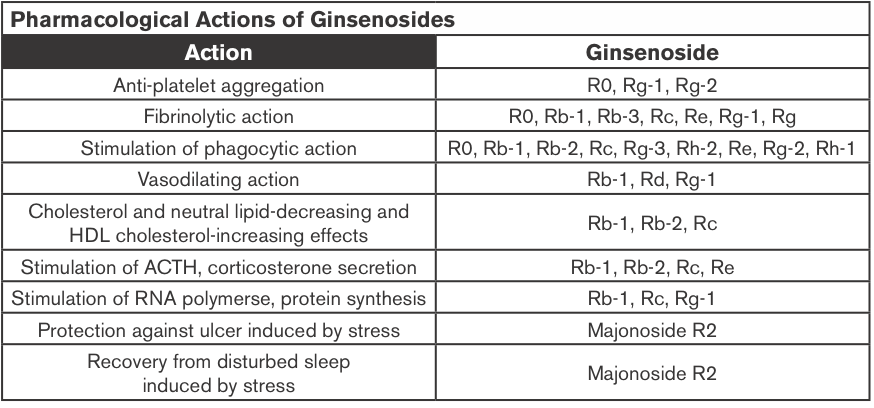
Ginsenoside content can vary widely depending on species, location of growth, and growing time before harvest. The root, the organ most often used, contains saponin complexes. These are often split into two groups: the Rb1 group (characterized by the protopanaxadiol presence: Rb1, Rb2, Rc and Rd) and the Rg1 group (protopanaxatriol: Rg1, Re, Rf, and Rg2). The potential health effects of ginsenosides include anti-carcinogenic, immunomodulatory, anti-inflammatory, anti-allergic, anti-atherosclerotic, anti-hypertensive, and anti-diabetic effects as well as anti-stress activity and effects on the central nervous system (Christensen, 2009).

Ginsenosides are considered the major pharmacologically active constituents, and approximately 12 types of ginsenosides have been isolated and structurally identified. Ginsenoside Rg3 was metabolized to ginsenoside Rh2 and protopanaxadiol by human fecal microflora (Bae et al., 2002). Ginsenoside Rg3 and the resulting metabolites exhibited potent cytotoxicity against tumor cell lines (Bae et al., 2002).

Screen-Shot-2014-03-28-at-11.53.41-am1

Ginseng Extracts (GE); Methanol-(alc-GE) or Water-extracted (w-GE) and ER+ Breast Cancer

Ginseng root extracts and the biologically active ginsenosides have been shown to inhibit proliferation of human cancer cell lines, including breast cancer. However, there are conflicting data that suggest that ginseng extracts (GEs) may or may not have estrogenic action, which might be contraindicated in individuals with estrogen-dependent cancers. The current study was designed to address the hypothesis that the extraction method of American ginseng (Panax quinquefolium) root will dictate its ability to produce an estrogenic response using the estrogen receptor (ER)-positive MCF-7 human breast cancer cell model. MCF-7 cells were treated with a wide concentration range of either methanol-(alc-GE) or water-extracted (w-GE) ginseng root for 6 days.

An increase in MCF-7 cell proliferation by GE indicated potential estrogenicity. This was confirmed by blocking GE-induced MCF-7 cell proliferation with ER antagonists ICI 182,780 (1 nM) and 4-hydroxytamoxifen (0.1 microM). Furthermore, the ability of GE to bind ERalpha or ERbeta and stimulate estrogen-responsive genes was examined. Alc-GE, but not w-GE, was able to increase MCF-7 cell proliferation at low concentrations (5-100 microg/mL) when cells were maintained under low-estrogen conditions. The stimulatory effect of alc-GE on MCF-7 cell proliferation was blocked by the ER antagonists ICI 182,780 or 4-hydroxyta-moxifen. At higher concentrations of GE, both extracts inhibited MCF-7 and ER-negative MDA-MB-231 cell proliferation regardless of media conditions.

These data indicate that low concentrations of alc-GE, but not w-GE, elicit estrogenic effects, as evidenced by increased MCF-7 cell proliferation, in a manner antagonized by ER antagonists, interactions of alc-GE with estrogen receptors, and increased expression of estrogen-responsive genes by alc-GE. Thus, discrepant results between different laboratories may be due to the type of GE being analyzed for estrogenic activity (King et al., 2006).

Anti-cancer

Previous studies suggested that American ginseng and notoginseng possess anti-cancer activities. Using a special heat-preparation or steaming process, the content of Rg3, a previously identified anti-cancer ginsenoside, increased significantly and became the main constituent in the steamed American ginseng. As expected, using the steamed extract, anti-cancer activity increased significantly. Notoginseng has a very distinct saponin profile compared to that of American ginseng. Steaming treatment of notoginseng also significantly increased anti-cancer effect (Wang et al., 2008).

Steam Extraction; Colorectal Cancer

After steaming treatment of American ginseng berries (100-120 ¡C for 1 h, and 120 ¡C for 0.5-4 h), the content of seven ginsenosides, Rg1, Re, Rb1, Rc, Rb2, Rb3, and Rd, decreased; the content of five ginsenosides, Rh1, Rg2, 20R-Rg2, Rg3, and Rh2, increased. Rg3, a previously identified anti-cancer ginsenoside, increased significantly. Two h of steaming at 120 ¡C increased the content of ginsenoside Rg3 to a greater degree than other tested ginsenosides. When human colorectal cancer cells were treated with 0.5 mg/mL steamed berry extract (120 ¡C 2 hours), the anti-proliferation effects were 97.8% for HCT-116 and 99.6% for SW-480 cells.

After staining with Hoechst 33258, apoptotic cells increased significantly by treatment with steamed berry extract compared with unheated extracts. The steaming of American ginseng berries hence augments ginsenoside Rg3 content and increases the anti-proliferative effects on two human colorectal cancer cell lines (Wang et al., 2006).

Glioblastoma

The major active components in red ginseng consist of a variety of ginsenosides including Rg3, Rg5 and Rk1, each of which has different pharmacological activities. Among these, Rg3 has been reported to exert anti-cancer activities through inhibition of angiogenesis and cell proliferation.

It is essential to develop a greater understanding of this novel compound by investigating the effects of Rg3 on a human glioblastoma cell line and its molecular signaling mechanism. The mechanisms of apoptosis by ginsenoside Rg3 were related with the MEK signaling pathway and reactive oxygen species. These data suggest that ginsenoside Rg3 is a novel agent for the chemotherapy of GBM (Choi et al., 2013).

Colon Cancer; Chemotherapy

Rg3 can inhibit the activity of NF-kappaB, a key transcriptional factor constitutively activated in colon cancer that confers cancer cell resistance to chemotherapeutic agents. Compared to treatment with Rg3 or chemotherapy alone, combined treatment was more effective (i.e., there were synergistic effects) in the inhibition of cancer cell growth and induction of apoptosis and these effects were accompanied by significant inhibition of NF-kappaB activity.

NF-kappaB target gene expression of apoptotic cell death proteins (Bax, caspase-3, caspase-9) was significantly enhanced, but the expression of anti-apoptotic genes and cell proliferation marker genes (Bcl-2, inhibitor of apoptosis protein (IAP-1) and X chromosome IAP (XIAP), Cox-2, c-Fos, c-Jun and cyclin D1) was significantly inhibited by the combined treatment compared to Rg3 or docetaxel alone.

These results indicate that ginsenoside Rg3 inhibits NF-kappaB, and enhances the susceptibility of colon cancer cells to docetaxel and other chemotherapeutics. Thus, ginsenoside Rg3 could be useful as an anti-cancer or adjuvant anti-cancer agent (Kim et al., 2009).

Prostate Cancer; Chemo-sensitizer

Nuclear factor-kappa (NF-kappaB) is also constitutively activated in prostate cancer, and gives cancer cells resistance to chemotherapeutic agents. Rg3 has hence also been found to increase susceptibility of prostate (LNCaP and PC-3, DU145) cells against chemotherapeutics; prostate cancer cell growth as well as activation of NF-kappaB was examined. It has been found that a combination treatment of Rg3 (50 microM) with a conventional agent docetaxel (5 nM) was more effective in the inhibition of prostate cancer cell growth and induction of apoptosis as well as G(0)/G(1) arrest accompanied with the significant inhibition of NF-kappaB activity, than those by treatment of Rg3 or docetaxel alone.

The combination of Rg3 (50 microM) with cisplatin (10 microM) and doxorubicin (2 microM) was also more effective in the inhibition of prostate cancer cell growth and NF-kappaB activity than those by the treatment of Rg3 or chemotherapeutics alone. These results indicate that ginsenoside Rg3 inhibits NF-kappaB, and enhances the susceptibility of prostate cancer cells to docetaxel and other chemotherapeutics. Thus, ginsenoside Rg3 could be useful as an anti-cancer agent (Kim et al., 2010).

Colon Cancer

Ginsenosides may not only be useful in themselves, but also for their downstream metabolites. Compound K (20-O-( β -D-glucopyranosyl)-20(S)-protopanaxadiol) is an active metabolite of ginsenosides and induces apoptosis in various types of cancer cells. This study investigated the role of autophagy in compound K-induced cell death of human HCT-116 colon cancer cells. Compound K activated an autophagy pathway characterized by the accumulation of vesicles, the increased positive acridine orange-stained cells, the accumulation of LC3-II, and the elevation of autophagic flux.

Compound K-provoked autophagy was also linked to the generation of intracellular reactive oxygen species (ROS); both of these processes were mitigated by the pre-treatment of cells with the anti-oxidant N-acetylcysteine.   Moreover, compound K activated the c-Jun NH2-terminal kinase (JNK) signaling pathway, whereas down-regulation of JNK by its specific inhibitor SP600125 or by small interfering RNA against JNK attenuated autophagy-mediated cell death in response to compound K.

Notably, compound K-stimulated autophagy as well as apoptosis was induced by disrupting the interaction between Atg6 and Bcl-2. Taken together, these results indicate that the induction of autophagy and apoptosis by compound K is mediated through ROS generation and JNK activation in human colon cancer cells (Kim et al., 2013b).

Lung Cancer; SCC

Korea white ginseng (KWG) has been investigated for its chemo-preventive activity in a mouse lung SCC model. N-nitroso-trischloroethylurea (NTCU) was used to induce lung tumors in female Swiss mice, and KWG was given orally. KWG significantly reduced the percentage of lung SCCs from 26.5% in the control group to 9.1% in the KWG group and in the meantime, increased the percentage of normal bronchial and hyperplasia. KWG was also found to greatly reduce squamous cell lung tumor area from an average of 9.4% in control group to 1.5% in the KWG group.

High-performance liquid chromatography/mass spectrometry identified 10 ginsenosides from KWG extracts, Rb1 and Rd being the most abundant as detected in mouse blood and lung tissue. These results suggest that KWG could be a potential chemo-preventive agent for lung SCC (Pan et al., 2013).

Leukemia

Rg1 was found to significantly inhibit the proliferation of K562 cells in vitro and arrest the cells in G2/M phase. The percentage of positive cells stained by SA-beta-Gal was dramatically increased (P < 0.05) and the expression of cell senescence-related genes was up-regulated. The observation of ultrastructure showed cell volume increase, heterochromatin condensation and fragmentation, mitochondrial volume increase, and lysosomes increase in size and number. Rg1 can hence induce the senescence of leukemia cell line K562 and play an important role in regulating p53-p21-Rb, p16-Rb cell signaling pathway (Cai et al., 2012).

Leukemia, Lymphoma

It has been found that Rh2 inhibits the proliferation of human leukemia cells concentration- and time-dependently with an IC(50) of ~38 µM. Rh2 blocked cell-cycle progression at the G(1) phase in HL-60 leukemia and U937 lymphoma cells, and this was found to be accompanied by the down-regulations of cyclin-dependent kinase (CDK) 4, CDK6, cyclin D1, cyclin D2, cyclin D3 and cyclin E at the protein level. Treatment of HL-60 cells with Rh2 significantly increased transforming growth factor- β (TGF- β ) production, and co-treatment with TGF- β neutralizing antibody prevented the Rh2-induced down-regulations of CDK4 and CDK6, up-regulations of p21(CIP1/WAF1) and p27(KIP1) levels and the induction of differentiation. These results demonstrate that the Rh2-mediated G(1) arrest and the differentiation are closely linked to the regulation of TGF- β production in human leukemia cells (Chung et al., 2012).

NSCLC

Ginsenoside Rh2, one of the components in ginseng saponin, has been shown to have anti-proliferative effect on human NSCLC cells and is being studied as a therapeutic drug for NSCLC. MicroRNAs (miRNAs) are small, non-coding RNA molecules that play a key role in cancer progression and prevention.

A unique set of changes in the miRNA expression profile in response to Rh2 treatment in the human NSCLC cell line A549 has been identified using miRNA microarray analysis. These miRNAs are predicted to have several target genes related to angiogenesis, apoptosis, chromatic modification, cell proliferation and differentiation. Thus, these results may assist in the better understanding of the anti-cancer mechanism of Rh2 in NSCLC (An et al., 2012).

Ginsenoside Concentrations

Ginsenosides, the major chemical composition of Chinese white ginseng (Panax ginseng C. A. Meyer), can inhibit tumor, enhance body immune function, prevent neurodegeneration. The amount of ginsenosides in the equivalent extraction of the nanoscale Chinese white ginseng particles (NWGP) was 2.5 times more than that of microscale Chinese white ginseng particles (WGP), and the extractions from NWGP (1000 microg/ml) reached a high tumor inhibition of 64% exposed to human lung carcinoma cells (A549) and 74% exposed to human cervical cancer cells (Hela) after 72 hours. Thia work shows that the nanoscale Chinese WGP greatly improves the bioavailability of ginsenosides (Ji et al., 2012).

Chemotherapy Side-effects

Pre-treatment with American ginseng berry extract (AGBE), a herb with potent anti-oxidant capacity, and one of its active anti-oxidant constituents, ginsenoside Re, was examined for its ability to counter cisplatin-induced emesis using a rat pica model. In rats, exposure to emetic stimuli such as cisplatin causes significant kaolin (clay) intake, a phenomenon called pica. We therefore measured cisplatin-induced kaolin intake as an indicator of the emetic response.

Rats were pre-treated with vehicle, AGBE (dose range 50–150 mg/kg, IP) or ginsenoside Re (2 and 5 mg/kg, IP). Rats were treated with cisplatin (3 mg/kg, IP) 30 min later. Kaolin intake, food intake, and body weight were measured every 24 hours, for 120 hours.

A significant dose-response relationship was observed between increasing doses of pre-treatment with AGBE and reduction in cisplatin-induced pica. Kaolin intake was maximally attenuated by AGBE at a dose of 100 mg/kg. Food intake also improved significantly at this dose (P<0.05). pre-treatment ginsenoside (5 mg/kg) also decreased kaolin intake >P<0.05). In vitro studies demonstrated a concentration-response relationship between AGBE and its ability to scavenge superoxide and hydroxyl.

Pre-treatment with AGBE and its major constituent, Re, hence attenuated cisplatin-induced pica, and demonstrated potential for the treatment of chemotherapy-induced nausea and vomiting. Significant recovery of food intake further strengthens the conclusion that AGBE may exert an anti-nausea/anti-emetic effect (Mehendale et al., 2005).

MDR

Because ginsenosides are structurally similar to cholesterol, the effect of Rp1, a novel ginsenoside derivative, on drug resistance using drug-sensitive OVCAR-8 and drug-resistant NCI/ADR-RES and DXR cells. Rp1 treatment resulted in an accumulation of doxorubicin or rhodamine 123 by decreasing MDR-1 activity in doxorubicin-resistant cells. Rp1 synergistically induced cell death with actinomycin D in DXR cells. Rp1 appeared to redistribute lipid rafts and MDR-1 protein.

Rp1 reversed resistance to actinomycin D by decreasing MDR-1 protein levels and Src phosphorylation with modulation of lipid rafts. Addition of cholesterol attenuated Rp1-induced raft aggregation and MDR-1 redistribution. Rp1 and actinomycin D reduced Src activity, and overexpression of active Src decreased the synergistic effect of Rp1 with actinomycin D. Rp1-induced drug sensitization was also observed with several anti-cancer drugs, including doxorubicin. These data suggest that lipid raft-modulating agents can be used to inhibit MDR-1 activity and thus overcome drug resistance (Yun et al., 2013).

Hypersensitized MDR Breast Cancer Cells to Paclitaxel

The effects of Rh2 on various tumor-cell lines for its effects on cell proliferation, induction of apoptosis, and potential interaction with conventional chemotherapy agents were investigated. Jia et al., (2004) showed that Rh2 inhibited cell growth by G1 arrest at low concentrations and induced apoptosis at high concentrations in a variety of tumor-cell lines, possibly through activation of caspases. The apoptosis induced by Rh2 was mediated through glucocorticoid receptors. Most interestingly, Rh2 can act either additively or synergistically with chemotherapy drugs on cancer cells. Particularly, it hypersensitized multi-drug-resistant breast cancer cells to paclitaxel.

These results suggest that Rh2 possesses strong tumor-inhibiting properties, and potentially can be used in treatments for multi-drug-resistant cancers, especially when it is used in combination with conventional chemotherapy agents.

MDR; Leukemia, Fibroblast Carcinoma

It was previously reported that a red ginseng saponin, 20(S)-ginsenoside Rg3 could modulate MDR in vitro and extend the survival of mice implanted with ADR-resistant murine leukemia P388 cells. A cytotoxicity study revealed that 120 microM of Rg3 was cytotoxic against a multi-drug-resistant human fibroblast carcinoma cell line, KB V20C, but not against normal WI 38 cells in vitro. 20 microM Rg3 induced a significant increase in fluorescence anisotropy in KB V20C cells but not in the parental KB cells. These results clearly show that Rg3 decreases the membrane fluidity thereby blocking drug efflux (Kwon et al., 2008).

MDR

Ginsenoside Rb1 is a representative component of panaxadiol saponins, which belongs to dammarane-type tritepenoid saponins and mainly exists in family araliaceae. It has been reported that ginsenoside Rb1 has diverse biological activities. The research development in recent decades on its pharmacological effects of cardiovascular system, anti-senility, reversing multi-drug resistance of tumor cells, adjuvant anti-cancer chemotherapy, and promoting peripheral nerve regeneration have been established (Jia et al., 2008).

Enhances Cyclophosphamide

Cyclophosphamide, an alkylating agent, has been shown to possess various genotoxic and carcinogenic effects, however, it is still used extensively as an anti-tumor agent and immunosuppressant in the clinic. Previous reports reveal that cyclophosphamide is involved in some secondary neoplasms.

C57BL/6 mice bearing B16 melanoma and Lewis lung carcinoma cells were respectively used to estimate the anti-tumor activity in vivo. The results indicated that oral administration of Rh(2) (5, 10 and 20 mg/kg body weight) alone has no obvious anti-tumor activity and genotoxic effect in mice, while Rh(2) synergistically enhanced the anti-tumor activity of cyclophosphamide (40 mg/kg body weight) in a dose-dependent manner.

Rh(2) decreased the micronucleus formation in polychromatic erythrocytes and DNA strand breaks in white blood cells in a dose-dependent way. These results suggest that ginsenoside Rh(2) is able to enhance the anti-tumor activity and decrease the genotoxic effect of cyclophosphamide (Wang, Zheng, Liu, Li, & Zheng, 2006).

Down-regulates MMP-9, Anti-metastatic

The effects of the purified ginseng components, panaxadiol (PD) and panaxatriol (PT), were examined on the expression of matrix metalloproteinase-9 (MMP-9) in highly metastatic HT1080 human fibrosarcoma cell line. A significant down-regulation of MMP-9 by PD and PT was detected by Northern blot analysis; however, the expression of MMP-2 was not changed by treatment with PD and PT. The results of the in vitro invasion assay revealed that PD and PT reduced tumor cell invasion through a reconstituted basement membrane in the transwell chamber. Because of the similarity of chemical structure between PD, PT and dexamethasone (Dexa), a synthetic glucocorticoid, we investigated whether the down-regulation of MMP-9 by PD and PT were mediated by the nuclear translocation of glucocorticoid receptor (GR). Increased GR in the nucleus of HT1080 human fibrosarcoma cells treated by PD and PT was detected by immunocytochemistry.

Western blot and gel retardation assays confirmed the increase of GR in the nucleus after treatment with PD and PT. These results suggest that GR-induced down-regulation of MMP-9 by PD and PT contributes to reduce the invasive capacity of HT1080 cells (Park et al., 1999).

Enhances 5-FU; Colorectal Cancer

Panaxadiol (PD) is the purified sapogenin of ginseng saponins, which exhibit anti-tumor activity. The possible synergistic anti-cancer effects of PD and 5-FU on a human colorectal cancer cell line, HCT-116, have been investigated.

The significant suppression on HCT-116 cell proliferation was observed after treatment with PD (25 microM) for 24 and 48 hours. Panaxadiol (25 microM) markedly (P < 0.05) enhanced the anti-proliferative effects of 5-FU (5, 10, 20 microM) on HCT-116 cells compared to single treatment of 5-FU for 24 and 48 hours.

Flow cytometric analysis on DNA indicated that PD and 5-FU selectively arrested cell-cycle progression in the G1 phase and S phase (P < 0.01), respectively, compared to the control condition. Combination use of 5-FU with PD significantly (P < 0.001) increased cell-cycle arrest in the S phase compared to that treated by 5-FU alone.

The combination of 5-FU and PD significantly enhanced the percentage of apoptotic cells when compared with the corresponding cell groups treated by 5-FU alone (P < 0.001). Panaxadiol hence enhanced the anti-cancer effects of 5-FU on human colorectal cancer cells through the regulation of cell-cycle transition and the induction of apoptotic cells (Li et al., 2009).

Colorectal Cancer

The possible synergistic anti-cancer effects of Panaxadiol (PD) and Epigallocatechin gallate (EGCG), on human colorectal cancer cells and the potential role of apoptosis in the synergistic activities, have been investigated.

Cell growth was suppressed after treatment with PD (10 and 20   µm) for 48   h. When PD (10 and 20   µm) was combined with EGCG (10, 20, and 30   µm), significantly enhanced anti-proliferative effects were observed in both cell lines. Combining 20   µm of PD with 20 and 30   µm of EGCG significantly decreased S-phase fractions of cells. In the apoptotic assay, the combination of PD and EGCG significantly increased the percentage of apoptotic cells compared with PD alone (p   <   0.01).

Data from this study suggested that apoptosis might play an important role in the EGCG-enhanced anti-proliferative effects of PD on human colorectal cancer cells (Du et al., 2013).

Colorectal Cancer; Irinotecan

Cell cycle analysis demonstrated that combining irinotecan treatment with panaxadiol significantly increased the G1-phase fractions of cells, compared with irinotecan treatment alone. In apoptotic assays, the combination of panaxadiol and irinotecan significantly increased the percentage of apoptotic cells compared with irinotecan alone (P<0.01). Increased activity of caspase-3 and caspase-9 was observed after treating with panaxadiol and irinotecan.

Data from this study suggested that caspase-3- and caspase-9-mediated apoptosis may play an important role in the panaxadiol enhanced anti-proliferative effects of irinotecan on human colorectal cancer cells (Du et al., 2012).

Anti-inflammatory

Ginsenoside Re inhibited IKK- β phosphorylation and NF- κ B activation, as well as the expression of pro-inflammatory cytokines, TNF- α and IL-1 β , in LPS-stimulated peritoneal macrophages, but it did not inhibit them in TNF- α – or PG-stimulated peritoneal macrophages. Ginsenoside Re also inhibited IRAK-1 phosphorylation induced by LPS, as well as IRAK-1 and IRAK-4 degradations in LPS-stimulated peritoneal macrophages.

Orally administered ginsenoside Re significantly inhibited the expression of IL-1 β and TNF- α on LPS-induced systemic inflammation and TNBS-induced colitis in mice. Ginsenoside Re inhibited colon shortening and myeloperoxidase activity in TNBS-treated mice. Ginsenoside Re reversed the reduced expression of tight-junction-associated proteins ZO-1, claudin-1, and occludin. Ginsenoside Re (20 mg/kg) inhibited the activation of NF- κ B in TNBS-treated mice. On the basis of these findings, ginsenoside Re may ameliorate inflammation by inhibiting the binding of LPS to TLR4 on macrophages (Lee et al., 2012).

Induces Apoptosis

Compound K activated an autophagy pathway characterized by the accumulation of vesicles, the increased positive acridine orange-stained cells, the accumulation of LC3-II, and the elevation of autophagic flux. Compound K activated the c-Jun NH2-terminal kinase (JNK) signaling pathway, whereas down-regulation of JNK by its specific inhibitor SP600125 or by small interfering RNA against JNK attenuated autophagy-mediated cell death in response to compound K. Compound K also provoked apoptosis, as evidenced by an increased number of apoptotic bodies and sub-G1 hypodiploid cells, enhanced activation of caspase-3 and caspase-9, and modulation of Bcl-2 and Bcl-2-associated X protein expression (Kim et al., 2013b).

Lung Cancer

AD-1, a ginsenoside derivative, concentration-dependently reduces lung cancer cell viability without affecting normal human lung epithelial cell viability. In A549 and H292 lung cancer cells, AD-1 induces G0/G1 cell-cycle arrest, apoptosis and ROS production. The apoptosis can be attenuated by a ROS scavenger – N-acetylcysteine (NAC). In addition, AD-1 up-regulates the expression of p38 and ERK phosphorylation. Addition of a p38 inhibitor, SB203580, suppresses the AD-1-induced decrease in cell viability. Furthermore, genetic silencing of p38 attenuates the expression of p38 and decreases the AD-1-induced apoptosis.

These data support development of AD-1 as a potential agent for lung cancer therapy (Zhang et al., 2013).

Pediatric AML

In this study, Chen et al. (2013) demonstrated that compound K, a major ginsenoside metabolite, inhibited the growth of the clinically relevant pediatric AML cell lines in a time- and dose-dependent manner. This growth-inhibitory effect was attributable to suppression of DNA synthesis during cell proliferation and the induction of apoptosis was accompanied by DNA double strand breaks. Findings suggest that as a low toxic natural reagent, compound K could be a potential drug for pediatric AML intervention and to improve the outcome of pediatric AML treatment.

Melanoma

Jeong et al. (2013) isolated 12 ginsenoside compounds from leaves of Panax ginseng and tested them in B16 melanoma cells. It significantly reduced melanin content and tyrosinase activity under alpha-melanocyte stimulating hormone- and forskolin-stimulated conditions. It significantly reduced the cyclic AMP (cAMP) level in B16 melanoma cells, and this might be responsible for the regulation down of MITF and tyrosinase. Phosphorylation of a downstream molecule, a cAMP response-element binding protein, was significantly decreased according to Western blotting and immunofluorescence assay. These data suggest that A-Rh4 has an anti-melanogenic effect via the protein kinase A pathway.

Leukemia

Rg1 can significantly inhibit the proliferation of leukemia cell line K562 in vitro and arrest the cells in G2/M phase. The percentage of positive cells stained by SA-beta-Gal was dramatically increased (P < 0.05) and the expression of cell senescence-related genes was up-regulated. The observation of ultrastructure showed cell volume increase, heterochromatin condensation and fragmentation, mitochondrial volume increase, and lysosomes increase in size and number (Cai et al., 2012).

Ginsenosides and CYP 450 Enzymes

In vitro experiments have shown that both crude ginseng extract and total saponins at high concentrations (.2000 mg/ml) inhibited CYP2E1 activity in mouse and human microsomes (Nguyen et al., 2000). Henderson et al. (1999) reported the effects of seven ginsenosides and two eleutherosides (active components of the ginseng root) on the catalytic activity of a panel of cDNA-expressed CYP isoforms (CYP1A2, CYP2C9, CYP2C19, CYP2D6, and CYP3A4) using 96-well plate fluorometrical assay.

Of the constituents tested, Ginsenoside Rd caused weak inhibitory activity against CYP3A4, CYP2D6, CYP2C19,and CYP2C9, but ginsenoside Re and ginsenoside Rf (200 mM) produced a 70% and 54%increase in the activity of CYP2C9 and CYP3A4, respectively. The authors suggested that the activating effects of ginsenosides on CYP2C9 and CYP3A4 might be due to a matrix effect caused by the test compound fluorescing at the same wavelength as the metabolite of the marker substrates. Chang et al. (2002) reported the effects of two types of ginseng extract and ginsenosides (Rb1, Rb2, Rc, Rd, Re, Rf, and Rg1) on CYP1 catalytic activities.

The ginseng extracts inhibited human recombinant CYP1A1, CYP1A2, and CYP1B1 activities in a concentration-dependent manner. Rb1, Rb2, Rc, Rd, Re, Rf, and Rg1 at low concentrations had no effect on CYP1 activities, but Rb1, Rb2, Rc, Rd, and Rf at a higher ginsenoside concentration (50 mg/ml) inhibited these activities. These results indicated that various ginseng extracts and ginsenosides inhibited CYP1 activity in an enzyme-selective and extract-specific manner (Zhou et al., 2003).

References

An IS, An S, Kwon KJ, Kim YJ, Bae S. (2012). Ginsenoside Rh2 mediates changes in the microRNA expression profile of human non-small-cell lung cancer A549 cells. Oncol Rep, 29(2):523-8. doi: 10.3892/or.2012.2136.


Bae EA, Han MJ, Choo MK et al. (2002). Metabolism of 20(S)- and 20(R)-ginsenoside R-g3 by human intestinal bacteria and its relation to in vitro biological activities. Biol. Pharm. Bull, 25:58–63.


Cai S, Zhou Y, Liu J, et al. (2012). Experimental study on human leukemia cell line K562 senescence induced by ginsenoside Rg1. Zhongguo Zhong Yao Za Zhi, 37(16):2424-8.


Cao M, Yu HS, Song XB, Ma BP. (2012) Advances in the study of derivatization of ginsenosides and their anti-tumor structure-activity relationship. Yao Xue Xue Bao, 47(7):836-43.


Chang TKH, Chen J, Benetton SA et al. (2002). In vitro effect of standardized ginseng extracts and individual ginsenosides on the catalytic activity of human CYP1A1, CYP1A2, and CYP1B1. Drug Metab. Dispos, 30:378–384.


Chen Y, Xu Y, Zhu Y, Li X. (2013). Anti-cancer effects of ginsenoside compound k on pediatric acute myeloid leukemia cells. Cancer Cell Int, 13(1):24. doi: 10.1186/1475-2867-13-24.


Choi YJ, Lee HJ, Kang DW, et al. (2013). Ginsenoside Rg3 induces apoptosis in the U87MG human glioblastoma cell line through the MEK signaling pathway and reactive oxygen species. Oncol Rep, 30(3): 1362-1370. doi: 10.3892/or.2013.2555.


Christensen LP. (2009). Ginsenosides chemistry, biosynthesis, analysis, and potential health effects. Adv Food Nutr Res., 55:1-99. doi: 10.1016/S1043-4526(08)00401-4.


Chung KS, Cho SH, Shin JS, et al. (2013). Ginsenoside Rh2 induces Cell-cycle arrest and differentiation in human leukemia cells by upregulating TGF- β expression. Carcinogenesis, 34(2):331-40. doi: 10.1093/carcin/bgs341.


Du GJ, Wang CZ, Zhang ZY, et al. (2012) Caspase-mediated pro-apoptotic interaction of panaxadiol and irinotecan in human colorectal cancer cells. J Pharm Pharmacol, 64(5):727-34. doi: 10.1111/j.2042-7158.2012.01463.x.


Du GJ, Wang CZ, Qi LW, et al. (2013). The synergistic apoptotic interaction of panaxadiol and epigallocatechin gallate in human colorectal cancer cells. Phytother Res, 27(2):272-7. doi: 10.1002/ptr.4707.


Henderson GL, Harkey MR, Gershwin, ME, et al. (1999). Effects of ginseng components on c-DNA-expressed cytochrome P450 enzyme catalytic activity. Life Sci, PL209–PL214.


Jeong YM, Oh WK, Tran TL, et al. (2013). Aglycone of Rh4 inhibits melanin synthesis in B16 melanoma cells: possible involvement of the protein kinase A pathway. Biosci Biotechnol Biochem, 77(1):119-25.


Ji Y, Rao Z, Cui J, et al. (2012). Ginsenosides extracted from nanoscale Chinese white ginseng enhances anti-cancer effect. J Nanosci Nanotechnol, 12(8):6163-7.


Jia WW, Bu X, Philips D, et al. (2004). Rh2, a compound extracted from ginseng, hypersensitizes Multi-drug-resistant tumor cells to chemotherapy. Can J Physiol Pharmacol, 82(7):431-7.


Jia JM, Wang ZQ, Wu LJ, Wu YL. (2008). Advance of pharmacological study on ginsenoside Rb1. Zhongguo Zhong Yao Za Zhi, 33(12):1371-7.


Kim YJ, Yamabe N, Choi P, et al. (2013a) Efficient Thermal Deglycosylation of Ginsenoside Rd and Its Contribution to the Improved Anti-cancer Activity of Ginseng. J Agric Food Chem.


Kim AD, Kang KA, Kim HS, et al. (2013b). A ginseng metabolite, compound K, induces autophagy and apoptosis via generation of reactive oxygen species and activation of JNK in human colon cancer cells. Cell Death Dis, 4:e750. doi: 10.1038/cddis.2013.273.


Kim SM, Lee SY, Cho JS, et al. (2010). Combination of ginsenoside Rg3 with docetaxel enhances the susceptibility of prostate cancer cells via inhibition of NF-kappaB. Eur J Pharmacol, 631(1-3):1-9. doi: 10.1016/j.ejphar.2009.12.018.


Kim SM, Lee SY, Yuk DY, et al. (2009). Inhibition of NF-kappaB by ginsenoside Rg3 enhances the susceptibility of colon cancer cells to docetaxel. Arch Pharm Res, 32:755–765. doi: 10.1007/s12272-009-1515-4.


King ML, Adler SR, Murphy LL. (2006). Extraction-dependent effects of American ginseng (Panax quinquefolium) on human breast cancer cell proliferation and estrogen receptor activation. Integr Cancer Ther, 5(3):236-43.


Kwon HY, Kim EH, Kim SW, et al. (2008). Selective toxicity of ginsenoside Rg3 on Multi-drug-resistant cells by membrane fluidity modulation. Arch Pharm Res, 31(2):171-7.


Lee IA, Hyam SR, Jang SE, Han MJ, Kim DH. (2012). Ginsenoside Re ameliorates inflammation by inhibiting the binding of lipopolysaccharide to TLR4 on macrophages. J Agric Food Chem, 60(38):9595-602.


Li XL, Wang CZ, Mehendale SR, et al. (2009). Panaxadiol, a purified ginseng component, enhances the anti-cancer effects of 5-fluorouracil in human colorectal cancer cells. Cancer Chemother Pharmacol, 64(6):1097-104. doi: 10.1007/s00280-009-0966-0.


Mehendale S, Aung H, Wang A, et al. (2005). American ginseng berry extract and ginsenoside Re attenuate cisplatin-induced kaolin intake in rats. Cancer Chemotherapy and Pharmacology, 56(1):63-9. doi: 10.1007/s00280-004-0956-1.


Nguyen TD, Villard PH, Barlatier A et al. (2000). Panax vietnamensis protects mice against carbon tetrachloride-induced hepatotoxicity without any modification of CYP2E1 gene expression. Planta Med, 66:714–719.


Pan J, Zhang Q, Li K, et al. (2013). Chemoprevention of lung squamous cell carcinoma by ginseng. Cancer Prev Res (Phila), 6(6):530-9. doi: 10.1158/1940-6207.CAPR-12-0366.


Park MT, Cha HJ, Jeong JW, et al. (1999). Glucocorticoid receptor-induced down-regulation of MMP-9 by ginseng components, PD and PT contributes to inhibition of the invasive capacity of HT1080 human fibrosarcoma cells. Mol Cells, 9(5):476-83.


Wang CZ and Yuan CS. (2008). Potential Role of Ginseng in the Treatment of Colorectal Cancer. Am. J. Chin. Med, 36:1019. doi: 10.1142/S0192415X08006545


Wang Z, Zheng Q, Liu K, Li G, Zheng R. (2006). Ginsenoside Rh(2) enhances anti-tumor activity and decreases genotoxic effect of cyclophosphamide. Basic Clin Pharmacol Toxicol, 98(4):411-5.


Wang CZ, Zhang B, Song WX, et al. (2006). Steamed American ginseng berry: ginsenoside analyzes and anti-cancer activities. Journal of agricultural and food chemistry, 54(26):9936-42.


Yun UJ, Lee JH, Koo KH, et al. (2013). Lipid raft modulation by Rp1 reverses Multi-drug resistance via inactivating MDR-1 and Src inhibition. Biochem Pharmacol, 85(10):1441-53. doi: 10.1016/j.bcp.2013.02.025.


Zhang LH, Jia YL, Lin XX, et al. (2013). AD-1, a novel ginsenoside derivative, shows anti-lung cancer activity via activation of p38 MAPK pathway and generation of reactive oxygen species. Biochim Biophys Acta, 1830(8):4148-59. doi: 10.1016/j.bbagen.2013.04.008.


Zhou Sf, Gao Yh, Jiang Wq et al. (2003) Interactions of Herbs with Cytochrome P450. DRUG METABOLISM REVIEWS, 35(1):35–98.

Xi Shu (Fr. seu Rx Camptothecae Acuminatae)

• Camptothecinum injection: 5 mg IM, bid or 15-20 mg added in normal saline 20 ml IV, once every other week, 200-250 mg as one course; 20 mg bladder perfusion twice a week or 5-10 mg intracancerous injection qod for bladder cancer. Camptothecinum suspension: 2.5-5.0 mg in 50% glucose 40 ml IV once every week, 50-100mg as one course, maintenance dose 2.5 mg every week, for liver cancer, leukemia and lymphoma. Hydroxycamptothecimum injection: 4-10 mg added in normal saline 20 ml IV qd or qod, 60-120mg as one course.

• Indications: bladder cancer, liver cancer, leukemia and lymphoma as indicated above.

• Pharmaceutical actions: Hydroxycamptothecine is an anti-carcinogenic which inhibits polymerase of DNA or damages DNA directly.

San Jiang Shan

(Ramulus et Folium Cephalotaxi Fortunei)

• Harringtonine injection: 1-6 mg added in 5-10% glucose 500 ml IV for drips daily, 5-7 days as one course, 1-3 courses may be applied at an 1-2 week intervals; homo-harringtonine, same dosage as harringtonine.

• Indications: this herb is mainly used as an anti-carcinogenic for acute non-lymphocytic leukemia such as acute myelocytic leukemia, acute monocytic leukemia, malignant lymphoma.

• Pharmacological action: 1) Harringtonine, homo-harringtonine, iso-harringtonine and deoxy-harringtonine are the anti-carcinogenic components, which interfere with the cell growth and mitotic activity, damage and inhibit the growth of S-stage cells and inhibit the synthesis of DNA. Harringtonine and homo-harringtonine are more effective for the interference of mitotic activity. 2) Inhibiting the hematopoeitic cells in bone marrow. 3) Causing irritant effect on digestive tract.