Category Archives: Bladder cancer

Anti-Bladder-Tumor Effect of Baicalein from Scutellaria baicalensis Georgi and Its Application In Vivo

Cancer: Bladder

Action: reduces cell growth and inhibits cell invasion

Evidence-Based Complementary and Alternative Medicine
Volume 2013 (2013), Article ID 579751, 12 pages
http://dx.doi.org/10.1155/2013/579751

Research Article

Jin-Yi Wu,1 Kun-Wei Tsai,2 Yi-Zhen Li,1 Yi-Sheng Chang,1 Yi-Chien Lai,1 Yu-Han Laio,1 Jiann-Der Wu,3 and Yi-Wen Liu1
1Department of Microbiology, Immunology and Biopharmaceuticals, College of Life Sciences, National Chiayi University, No. 300 Syuefu Road, Chiayi 600, Taiwan
2Department of Internal Medicine, Buddhist Tzuchi Dalin General Hospital, Dalin Town, Chiayi 622, Taiwan
3Department of Pathology, Chiayi Christian Hospital, Chiayi 600, Taiwan

Received 16 November 2012; Revised 22 January 2013; Accepted 5 February 2013

Academic Editor: Jen-Hwey Chiu

Copyright © 2013 Jin-Yi Wu et al. This is an open access article distributed under the Creative Commons Attribution License, which permits unrestricted use, distribution, and reproduction in any medium, provided the original work is properly cited.

Abstract

Some phytochemicals with the characteristics of cytotoxicity and/or antimetastasis have generated intense interest among the anticancer studies. In this study, a natural flavonoid baicalein was evaluated in bladder cancer in vitro and in vivo. Baicalein inhibits 5637 cell proliferation. It arrests cells in G1 phase at 100 μM and in S phase below 75 μM. The protein expression of cyclin B1 and cyclin D1 is reduced by baicalein. Baicalein-induced p-ERK plays a minor role in cyclin B1 reduction. Baicalein-inhibited p65NF-κB results in reduction of cell growth. Baicalein-induced pGSK(ser9) has a little effect in increasing cyclin B1/D1 expression instead. The translation inhibitor cycloheximide blocks baicalein-reduced cyclin B1, suggesting that the reduction is caused by protein synthesis inhibition. On the other hand, neither cycloheximide nor proteasome inhibitor MG132 completely blocks baicalein-reduced cyclin D1, suggesting that baicalein reduces cyclin D1 through protein synthesis inhibition and proteasomal degradation activation. In addition, baicalein also inhibits cell invasion by inhibiting MMP-2 and MMP-9 mRNA expression and activity. In mouse orthotopic bladder tumor model, baicalein slightly reduces tumor size but with some hepatic toxicity. In summary, these results demonstrate the anti-bladder-tumor properties of the natural compound baicalein which shows a slight anti-bladder-tumor effect in vivo.

1. Introduction

Bladder cancer is the seventh most common type of cancer in worldwide man [1] and fourth in man of United States [2]. More than 90% of bladder cancers are transitional cell carcinoma (TCC), and approximately 80% of TCC belong to noninvasive papillary carcinoma that is a low-grade intraurothelial neoplasia with high recurrence. The other 20% of TCCs initiated from carcinoma in situ are at a high risk of processing to muscle invasive disease with a substantial risk for the development of distant metastasis [3, 4]. More than 10% of the low-grade papillary tumors eventually progress to high-grade muscle invasive bladder tumors. Most of the deaths from bladder cancer patients are due to invasive cancer metastasis [5], which has been a leading problem in the cancer therapy field. Multiple drugs chemotherapy has been applied for the therapy of metastatic bladder cancer; however, the adverse effect and resistance usually limit its clinical result. Therefore, some phytochemicals with the characteristics of cytotoxicity and/or antimetastasis have generated intense interest among the anticancer studies.

Baicalein, one of four major flavonoids existed in the root of Scutellaria baicalensis Georgi, has excellent antioxidant and anti-inflammatory activities [6, 7]. In traditional Chinese herb medicine, the root of Scutellaria baicalensis Georgi was usually gathered before Tomb-Sweeping Day and decocted for the purpose of “cleansing heart” and “removing toxins,” for example, cough with yellow sputum, jaundice, swelling and pain of eye, and so on. Wogonin, another one of the major flavonoids in the root of Scutellaria baicalensis Georgi, has been reported to reduce inflammatory cyclooxygenase-2 expression by c-Jun inhibition [8]. In addition to the anti-inflammatory effect of wogonin, baicalein has been reported to apply in cancer therapy by its cytotoxicity [9–11] and its anti-metastasis activity [12–14] recently. In human pancreatic cancer cells, 15~50 μM baicalein induces apoptotic cell death through downregulation of an antiapoptotic protein Mcl-1 [11]. In human bladder cancer cells, 60~80 μM baicalein retards cell growth by inhibiting CDC2 kinase activity [9]. Sixty μM baicalein also induces bladder cancer cells death, but baicalein-induced p-Akt and -H2AX expression plays a protective role against cell death [10]. Moreover, 10~50 μM baicalein inhibits cell migration and invasion through inhibiting MMP-2/9 activity in human hepatoma cells [12] and human breast cancer cells [13]. In human skin carcinoma, 40 μM baicalein inhibits cell invasion through inhibiting an anchor protein Ezrin expression [14]. Recently, baicalein is proven to be genotoxic without producing chromosomal alterations and mutagenesis which results in the severe side effect in cancer chemotherapy [15]. According to the above data, baicalein is a candidate worth development in anticancer therapy.

In this study, the anticancer effect of baicalein was analyzed in bladder cancer cells in vitro and in an orthotopic bladder tumor model in vivo. In vitro, the correlation of baicalein-induced change in Akt, ERK, p38, and p65NF-κB pathways and cell viability was analyzed. In vivo, the antitumor effect and renal and hepatic toxicities were evaluated.

2. Materials and Methods

2.1. Cell Culture and Drug Preparation

Human bladder papillary transitional cell carcinoma 5637 cells were obtained from the Bioresource Collection and Research Center (Hsinchu, Taiwan). Mouse bladder carcinoma MB49 cells were kindly provided by Dr. Timonthy L. Ratliff (Purdue Cancer Center, West Lafayette, IN, USA). 5637 and MB49 cells were maintained in RPMI 1640 medium supplied with 10% fetal bovine serum (FBS), 1% penicillin, and 1% streptomycin. Cells were incubated in a CO2 incubator at 37°C, with 5%  CO2 and 95% filtered air. Baicalein was isolated from the root of Scutellaria baicalensis Georgi, identified [16] and dissolved in DMSO. For culture cell assay, baicalein was added in culture medium containing 0.1% DMSO. For mouse assay, baicalein was intraperitoneally injected in mice containing 10% DMSO and 90% propylene glycol (0.8 mg/100 μL/mouse).

2.2. Reagents and Antibodies

3-(4,5-Dimethylthiazol-2-yl)-2,5-diphenyltetrazolium bromide (MTT), propidium iodide (PI), ribonuclease A (RNase A), propylene glycol, MG-132, and crystal violet were purchased from Sigma (St. Louis, MO, USA). Anti-phospho-AKT(thr308), anti-phospho-AKT(ser473), and anti-cyclin D1 antibodies were purchased from Santa Cruz (Santa Cruz, CA, USA). Anti-α-tubulin, anti-β-actin, and anti-phospho-GSK-3β(ser9) antibodies were purchased from GeneTex (Taichung, Taiwan). Anti-cyclin B1 was purchased from Epitomics (Burlingame, CA, USA). Anti-Bub3 was purchased from BD Biosciences (San Jose, CA, USA). Anti-p65NF-κB, anti-phospho-ERK(thr202/tyr204), and anti-phospho-p38(thr180/tyr182) were purchased from Cell Signaling Technology (Danvers, MA, USA). The Millicell Hanging Cell Culture Inserts of Transwell system was purchased from Millipore (Billerica, MA, USA). Peroxidase-conjugated secondary antibodies were purchased from Jackson ImmunoResearch (West Grove, PA, USA).

2.3. Cell Viability Assay

Cell number was determined by colorimetric MTT assay. 5637 cells were cultured in 24-well plates at a density of 5 × 104 cells/well. After 24 h, cells were incubated with various concentrations of baicalein or 0.1% DMSO for another 24~72 h. Then MTT was added into medium for 2 h, the medium was discarded, and DMSO was added to dissolve the formazan product. Each well was measured by light absorbance at 490 nm. The result was expressed as a percentage, relative to 0.1% DMSO-treated control group.

2.4. Cell Cycle Analysis

Around 2 × 106 5637 cells were seeded in 100 mm dishes. After 24 h incubation for attachment, baicalein or DMSO was added. After baicalein treatment for 24 h and 48 h, cells were trypsinised, centrifuged, and fixed with ice-cold 75% ethanol overnight at 4°C. After removing the ethanol, cells were stained with a DNA staining solution (containing 1 mg/mL PI and 10 mg/mL RNase A dissolved in PBS) for 30 min at room temperature. The DNA content of the stained cells was measured using a FACScan flow cytometer. The cell doublets were removed by gating the left area of FL2-W/FL2-A plot for analysis. Cell cycle data from flow cytometry was analysed using ModFit LT software.

2.5. Cell Migration Assay

5637 cells were seeded in 6-well plates. After cells had reached confluence, a wound was made by a 200 μL plastic tip in each well. The wells were then washed twice with PBS to remove cell debris and then incubated with culture medium with DMSO (control) or baicalein. After 24 h incubation, each well was photographed by a phase contrast microscopy. The empty area was calculated by computer, and the cell mobility was calculated by (scratch area − empty area of baicalein treatment) × 100%/(scratch area − empty area of control). Measurements were performed in triplicate and presented as mean ± SE from three independent experiments.

2.6. Cell Invasion Assay

The invasion assay was analyzed using a Matrigel (BD Biosciences)-coated Transwell system (Millipore). The upper chamber of the transwell was coated with 25 μg Matrigel. 5637 cells (1 × 105) in serum-free RPMI-1640 media were seeded onto Matrigel-coated Transwell. The upper and lower chamber media were added baicalein or 0.1% DMSO. In the lower chambers, 10% FBS was added as a chemoattractant. After a 24 h incubation time, the cells that remained on the upper surface of the filter membrane were removed, and the cells on the opposite surface of the filter membrane were stained with 4% paraformaldehyde for 30 s and photographed under microscopy at 200x magnification. The number of migrated cells was counted in five randomly chosen microscope fields.

2.7. RT-PCR

Total RNA was isolated from cells. Reverse transcription (RT) was performed on 2 μg of total RNA by 1.5 μM random hexamer and RevertAid reverse transcriptase (Fermentas); then 1/20 volume of reaction mixture was used for PCR with MMP-2 specific primers (5′CTTCCAAGTCTGGAGCGATGT3′, 5′TACCGTCAAAGGGGTATCCAT3′), MMP-9 specific primers (5′AAGATGCTGCTGTTCAGCGGG3′, 5′GTCCTCTGGGCACTGCAGGAT3′), and GAPDH specific primers (5′CGGATTTGGTCGTATTGG3′, 5′AGATGGTGATGGGATTTC3′). The PCR products were analyzed by 1% agarose gel.

2.8. Gelatin-Zymography Assay

The enzymatic activities of MMP-2 and MMP-9 were determined by gelatin-zymography. 3 × 106 cells were seeded in 10 cm dish for 24 h and then maintained in serum-free medium with various concentrations of baicalein. The conditioned medium was collected 24 h after drug treatment, concentrated by using an Amicon Ultracel YM-10 filter. Twenty micrograms of protein obtained from the concentrated medium was mixed with nonreducing sample buffer and subjected to electrophoresis (8% SDS-PAGE copolymerize with 0.1% gelatin as substrate). The gel was washed twice (15 min/time) with 2.5% Triton X-100 and incubated at 37°C for 16~20 h in 50 mM Tris-HCl (pH 7.8), 10 mM CaCl2, and 0.01% NaN3. The gel was stained with 0.15% Coomassie brilliant blue R-250 and destained in 50% methanol and 10% acetic acid until the gelatinolytic activities were detected as clear bands against a blue background.

2.9. Mouse Orthotopic Bladder Tumor Model

The female C57BL/6 mice aged five to six weeks were provided by the National Laboratory Animal Center (Taipei, Taiwan) and maintained at our animal care facility for one week prior to use. The implantation of murine bladder cancer cells MB49 into C57BL/6 mice was carried out similarly as previous report [17, 18].After MB49 inoculation (day 1), mice were randomly assigned to two groups (10 mice/group). One group was intraperitoneally treated with vehicle (10% DMSO and 90% propylene glycol), and the other group received 0.8 mg/mouse baicalein intraperitoneally for 9 times. At the 21th day, the mice were sacrificed and the bladder volumes were measured before formalin fixation. After cutting into 4 μm sections, the slides of each mouse were confirmed under a microscope in histology by hematoxylin and eosin staining. The experiment was approved by the Institutional Animal Care and Use Committee of National Chiayi University.

2.10. Statistical Analysis

The values shown are mean ± SEM. Data are statistically evaluated by one-way ANOVA of SigmaPlot 11.0 and shown significantly different in , , and .

3. Results

3.1. Cytotoxicity and Proliferation Inhibition of Baicalein in 5637 Bladder Cancer Cells

Cytotoxicity of baicalein was analyzed by MTT assay. The result shows that baicalein dose-dependently inhibits cell viability after 24 h treatment (Figure 1(a)). Below 50 μM, baicalein did not induce cell death because there were no floating cells after treatment. When the concentration reached 100 μM, baicalein causes 33% cell number down with dead floating cells in the culture medium. To distinguish the fact of cell death and proliferation inhibition, the direct cell count analysis was applied after baicalein treatment. The result of Figure 1(b) suggests that under 50 μM, baicalein does not reduce total cell number after treatment for 72 h. Only for the concentration higher than 75 μM, baicalein induces cell death dose-dependently. These data suggest that baicalein induces growth inhibition at a dose lower than 50 μM and causes cell death at a dose higher than 75 μM in 5637 cells.

fig1
Figure 1: Effect of baicalein on cell growth. (a) Cytotoxicity of baicalein in 5637 bladder cancer cells. 5637 cells were initially seeded at 1 × 105 cells per well in 24-well plates and then treated with various concentrations of baicalein or vehicle (0.1% DMSO) for 24 h. The cell viability was measured by MTT assay. Measurement is performed from three independent experiments ( compared with vehicle). (b) Baicalein dose-dependently inhibits cell growth of 5637 cells. Cells were initially seeded at 1 × 105 cells (day-1) per well in 24-well plates and then treated with various concentrations of baicalein or vehicle (0.1% DMSO) for 24~72 h. The cell number was counted by trypan blue dye exclusion assay. The dotted line indicates the cell number on day 0.
3.2. Baicalein Induces Cell Cycle Arrest and Decreases Cyclin B1/D1 Expression of 5637 Bladder Cancer Cells

The cell cycle distribution changed by baicalein was analyzed by flow cytometric assay. Baicalein arrests cells in S phase after 24 h treatment at the concentration under 75 μM and in G1 phase at 100 μM. After treatment for 48 h, 100 μM baicalein continued to arrest cells in G1 without sub-G1 formation (Figure 2(a)). It suggests that baicalein, less than 50 μM, caused S phase arrest without significant cytotoxicity. One hundred μM baicalein arrested cells in G1 phase and induced cytotoxicity. One of G1/S transition promotion factors, cyclin D1, was dose-dependently decreased by baicalein (Figure 2(b)). It may contribute the reason to 100 μM baicalein-induced G1 arrest. Baicalein also decreased cyclin B1 expression dose-dependently (Figure 2(b)). Because cyclin B1 is an essential factor for entering G2/M phase, baicalein-decreased cyclin B1 may lead to S phase arrest. Baicalein immediately decreases cyclin D1 expression after treatment for 2 h (Figure 2(b)). It suggests that 100 μM baicalein effectively and quickly inhibits cell cycle progression at G1 phase.
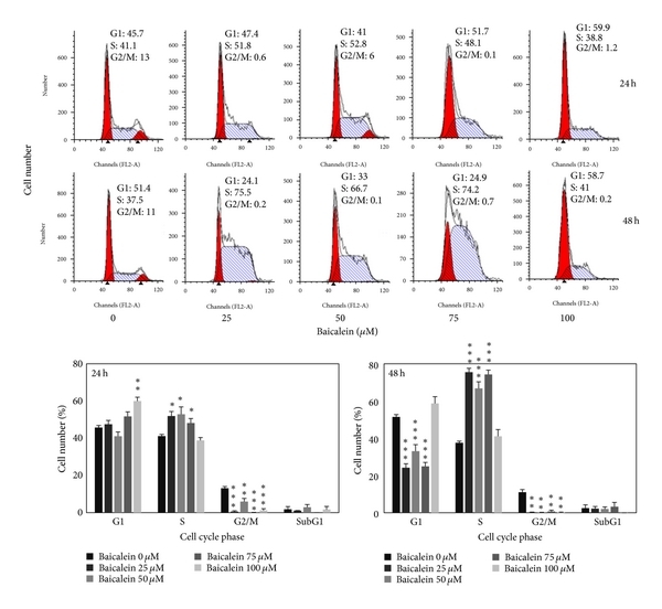
579751.fig.002afig2
Figure 2: Effect of baicalein on cell cycle phase distribution in 5637 cells. (a) Baicalein induces cell cycle arrest. Cells were treated with vehicle or baicalein for 24 and 48 h, then were collected for cell cycle analysis (, , compared with vehicle). (b) Effect of baicalein on cyclin B1/D1 expression. Left, cells were treated with baicalein for 24 h. Right, cells were treated with 100 μM baicalein for 2, 6, 12, and 24 h.
3.3. Effect of Baicalein on the Regulation of Upstream Signal Factors

The intracellular signal factors p-GSK3β(ser9), p-AKT(thr308), p-AKT(ser473), p-ERK, and p-p38 were analyzed after baicalein treatment. The result of Figure 3(a) indicates that baicalein increases the phosphorylation of GSK3β(ser9), ERK(thr202/tyr204), and p38(thr180/tyr182). Though baicalein decreased the phosphorylation of AKT(thr308) and did not change p-AKT(ser473), the downstream GSK3β(ser9) was still phosphorylated at 24 h treatment. The time course of these baicalein-induced changes was also analyzed. As shown in Figure 3(b), baicalein inhibited p-AKT(thr308) phosphorylation from 2 h to 24 h; it suggests that baicalein inhibits Akt activity. GSK3β(ser9) was phosphorylated by baicalein from 2 h to 24 h, suggesting that baicalein also inhibits GSK3β activity. Both ERK and p38 pathways were early activated from 2 h to 24 h after baicalein treatment, ERK especially. The effect of baicalein on p65NF-κB was also analyzed. Without extracellular stimulation, most of p65NF-κB was found in cytoplasmic fraction in 5637 cells (Figure 3(c)). Baicalein dose-dependently inhibited the nuclear protein expression of p65NF-κB (Figure 3(c)). In summary, baicalein inhibits AKT and GSK3β activities, activates ERK and p38 pathways, and inhibits p65NF-κB-driven signals.
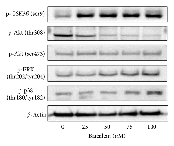
579751.fig.003afig3
Figure 3: Influence of baicalein on the signal pathways. (a) Effect of baicalein on the phosphorylation of GSK3β, AKT, ERK, and p38. 5637 cells were treated with 0.1% DMSO or baicalein for 24 h. (b) Time-course of baicalein-changed signal protein phosphorylation. 5637 cells treated with 100 μM baicalein or 0.1% DMSO for 2, 6, 12, and 24 h. The total cell lysates were extracted for western blot analysis. β-Actin was used as a loading control. (c) Effect of baicalein on the nuclear p65NF-κB expression. 5637 cells were treated with 0.1% DMSO or baicalein for 24 h. The cytoplasmic and nuclear extracts were prepared for Western blot analysis. α-Tubulin and Bub3 are the loading control of cytoplasmic and nuclear fraction, respectively.
3.4. Effect of Various Signal Protein Inhibitors on the Baicalein-Changed Cyclin B1/D1 Expression and Cell Viability

In order to understand the correlation between upstream signals and cyclin B1/D1 reduction, some specific inhibitors were used. Lithium chloride (LiCl) induces GSK3β(ser9) phosphorylation and inhibits GSK3βactivity [19, 20]. Baicalein or LiCl increased p-GSK3β(ser9), but only baicalein decreased cyclin B1/D1 expression (Figure 4(a)). It suggests that baicalein-inhibited cyclin B1/D1 expression is not mediated by GSK3βinhibition. On the contrary, LiCl dose-dependently increased cyclin B1/D1 expression, it suggests that baicalein-inhibited GSK3β pathway causes cyclin B1/D1 increase instead. LY294002, the inhibitor of PI3K-Akt pathway, inhibited the phosphorylation of AKT(ser473) but increased the phosphorylation of GSK3β(ser9) (Figure 4(b)). However, unlike baicalein, LY294002 did not reduce cyclin B1/D1 expression (Figure 4(b)). U0126, the inhibitors of MEK-ERK, slightly reversed baicalein-decreased cyclin B1 but not cyclin D1 (Figure 4(c)). The p38 kinase inhibitor SB203580 did not reverse baicalein-decreased cyclin B1/D1 expression (Figure 4(d)). Ro106-9920, an inhibitor of p65NF-κB, did not decrease cyclin B1/D1 expression (Figure 4(e)). These data indicate that baicalein-inhibited cyclin B1 is slightly mediated by ERK activation. The relationship of cell viability and baicalein-induced change in p-GSK3β(ser9), p-ERK, p-p38, and p65NF-κB was also analyzed. Using MTT assay (Figure 4(f)), MEK-ERK inhibitor U0126 and p38 kinase inhibitor SB203580 did not affect baicalein-reduced cell viability; the PI3K inhibitor LY294002 deteriorated baicalein-reduced cell viability; the p65NF-κB inhibitor Ro106-9920 reduced cell viability directly. It suggests that baicalein-induced p38, ERK, and GSK3β(ser9) phosphorylation does not play essential roles in cell growth inhibition. Only the baicalein-inhibited p65NF-κB activity leads to reduction of cell viability. In order to find out baicalein-reduced cyclin B1/D1 caused by de novo protein synthesis inhibition or proteasomal degradation stimulation, the translation inhibitor cycloheximide and the proteasome inhibitor MG132 were used for this study. After cyclohexamide treatment, baicalein did not reduce cyclin B1 anymore (Figure 4(g)). But baicalein still reduced cyclin D1 expression in the presence of cycloheximide or MG132 (Figure 4(g)). It suggests both de novo protein synthesis inhibition and proteasomal degradation stimulation are involved in baicalein-reduced cyclin D1 expression, and cyclin B1 decrease is only caused by de novo protein synthesis inhibition.
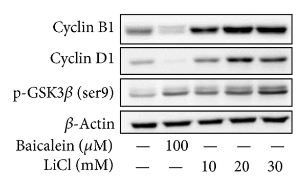
579751.fig.004afig4
Figure 4: Effect of various inhibitors on baicalein-reduced cyclin B1/D1 expression. (a–e) Effect of LiCl (a), LY294002 (b), U0126 (c), SB203580 (d), and Ro106-9920 (e) on baicalein-reduced cyclin B1/D1 expression. (f) Effect of various inhibitors on baicalein-inhibited cell viability. All above inhibitors were pretreated for 1 h and baicalein treatment for 24~72 h. The concentration of each chemicals: baicalein is 100 μM and others are 10 μM. (g) Effect of cycloheximide or MG132 on baicalein-reduced cyclin B1/D1 expression. Cycloheximide was pretreated for 30 min and baicalein treatment for 1 h in cyclin D1 detection and baicalein treatment for 6 h in cyclin B1 detection. MG132 was pretreated for 1 h and baicalein treatment for 6 h. The extracted cell lysates or nuclear proteins were analyzed by western blot.
3.5. Baicalein Blocks Migration and Invasion of 5637 Bladder Cancer Cells

Using scratch assay, baicalein dose-dependently inhibited cell migration (Figure 5(a)). At 100 μM, baicalein shows 60% inhibition in cell migration, which is more effective than the inhibition in cell viability (33% inhibition at 100 μM, Figure 1(a)). By matrigel-coated invasion assay, baicalein also shows a significant inhibition dose-dependently (Figure 5(b)). On the other hand, baicalein reduced MMP2 and MMP9 mRNA expression (Figure 5(c)) and enzymatic activity (Figure 5(d)) in 5637 cells. It suggests that the baicalein-inhibited MMP2/9 activity may contribute its anti-migration and anti-invasion activity.

579751.fig.005afig5
Figure 5: Anti-migration and anti-invasion activities of baicalein in 5637 cells. (a) Baicalein inhibits 5637 cell migration. Bottom chart is the percentage of migrated cells as control is 100%. (b) Baicalein inhibits 5637 cell invasion. Bottom chart is the percentage of invaded cells as control is 100%. (c) Effect of baicalein on the mRNA expression of MMP-2 and MMP-9. Bottom charts are the quantitative results from three independent experiments. (d) Effect of baicalein on the activities of MMP-2 and MMP-9. The conditioned medium was collected 24 h after drug treatment. Twenty micrograms of protein obtained from the concentrated medium was analyzed by gelatin-zymography assay. (, , compared with vehicle).
3.6. Baicalein Slightly Inhibits Tumor Growth with Some Hepatotoxicity in a Mouse Orthotopic Bladder Tumor Model In Vivo

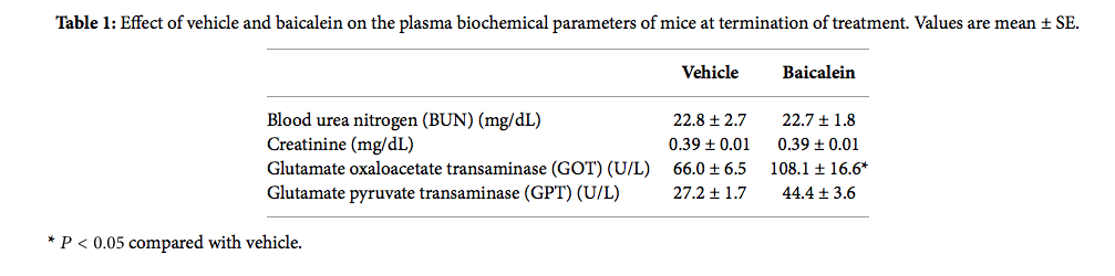
Based on the antigrowth and antimetastasis activity of baicalein in cell assay, the in vivo antitumor assay was analyzed. After bladder cell implantation on day 1, baicalein treatment started on day 8. The treatment did not show toxicity in appearance and body weight (Figure 6(a)). Baicalein did not significantly reduce bladder size, but the mean bladder volume was still reduced in baicalein-treated mice (from 49.5 mm3 to 35.9 mm3 in Figure 6(b)). The blood biochemical analysis shows no significant change in serum BUN and creatinine between control and baicalein treatment groups, a little increase in GPT value but without statistical significance, and a significant increase in serum GOT (Table 1). It suggests that baicalein treatment causes some hepatic toxicity in mice.

table1tab1
Table 1: Effect of vehicle and baicalein on the plasma biochemical parameters of mice at termination of treatment. Values are mean ± SE.
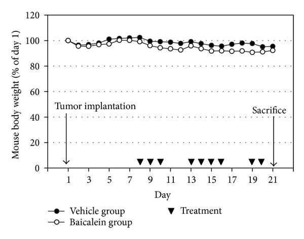
579751.fig.006afig6
Figure 6: The antitumor effect of baicalein in vivo. (a) Mouse body weight and drug schedule in the mouse orthotopic bladder tumor model. After MB49 cell implantation, baicalein or vehicle was applied by intraperitoneal injection. Mouse body weight was recorded every day. At the 21th day, the mice were sacrificed. (b) Effect of baicalein in reducing bladder tumor size. The bladder volume of each mouse was measured. There are 9 mice survived at the 21th day in vehicle group and 7 in baicalein group.
4. Discussion

This study provides some new information about baicalein used in the anticancer therapy. In cell study, baicalein decreases cyclin B1 protein expression through inhibiting de novo protein synthesis and inhibits cyclin D1 by inhibiting protein synthesis and promoting proteasomal degradation. Baicalein-inhibited cyclin B1 is partially mediated by ERK activation. Among the signal transduction molecules AKT, GSK3β, ERK, p38, and p65NF-κB, p65NF-κB inhibition plays the most important role in baicalein-reduced cell viability. In mouse orthotopic bladder tumor model, baicalein has a little inhibition effect on orthotopic bladder tumor growth but with some hepatic toxicity.

Baicalein produces different cytotoxicity in different cell lines. For example, it causes cell cycle arrest at G1 phase in breast cancer [21] and oral squamous cell carcinoma [22], at S phase in lung nonsmall carcinoma cell [23] and at G2/M phase in BFTC905 bladder cancer cells [9]. The differences may be caused by different doses and different cells used. In 5637 bladder cancer cells, baicalein arrests cells at S phase under 75 μM and at G1 phase at 100 μM without apoptotic cells (Figure 2(a)). In pancreatic carcinoma PaCa cells [11], bladder cancer BFTC905 cells [9], and colorectal carcinoma HCT116 cells [10], baicalein induces cell apoptotic death at the dose between 5 to 60 μM. Baicalein also has a wide range on cytotoxicity of different cell lines, the IC50 is under 20 μM in gastric cancer cells AGS and MKN-28 [24], prostate carcinoma LNCaP [25], and JCA-1 [26], between 20 to 50 μM in leukemia HL-60 [27], bladder cancer BFTC905 [9], hepatic cancer Hep G2 [28], and myeloma cell U266 [29], and more than 100 μM in 5637 bladder cancer (Figure 1(a)), oral squamous carcinoma HSC-3 [22], leukemia THP-1, and osteogenic cancer cell HOS [30]. Although the detail mechanisms about the wide-range cytotoxicity are still unclear, this property may provide a specific and lower hazard anticancer effect for the higher sensitive tumors.

PI3K, the upstream signal of AKT, has been reported to be inhibited by baicalein [31]. In our study, baicalein inhibits pAKT(thr308) phosphorylation and has no influence on pAKT(ser473) (Figure 2(b)). The pAKT inhibition phenomenon also has been reported in prostate cancer cell DU145 [32] and oral squamous carcinoma HSC-3 [22]. On the other hand, the pAKT(ser473) activation has been reported in bladder cancer BFTC905 [9, 10]. Because the pAKT(thr308) phosphorylation site is the direct target site for PI3K-PDK1 [33], it will be downregulated after PI3K inhibition by baicalein (Figure 4(b)). The ser473 site of AKT is phosphorylated by rictor-mTOR [34], not PI3K; therefore, it may be the reason for the no effect of baicalein on the phosphorylation of pAKT(ser473). Even though the pAKT(thr308) is decreased by baicalein, pGSK3β(ser9), one AKT downstream [35], is still phosphorylated by baicalein (Figure 3(b)). Because the phosphorylation of pGSK3β(ser9) is achieved by numerous kinases, not only AKT [35], baicalein-induced pGSK3β(ser9) may be caused by the influence of other kinase(s).

NF-κB, an important inflammatory transcription factor, is inhibited by baicalein in 5637 cells (Figure 3(c)). Baicalein-inhibited p65NF-κB activation has also been reported in human mast cells [36], mouse macrophages [37, 38], human myeloma cells [29], and brain microglia [39, 40]. According to the important role of p65NF-κB in tumor progression and metastasis [41, 42], the inhibition of baicalein on nuclear NF-κB is a critical function in its anti-inflammation and anticancer application. In human hepatoma cells, baicalein shows anti-migration property with NF-κB inhibition [12]. There is one report indicates that GSK3β inhibition results in inhibiting NF-κB activity [43]; therefore, the mechanism of baicalein-inhibited p65NF-κB activity may be partially mediated by baicalein-inhibited GSK3β. In Figure 4(f), among the 4 signal inhibitors, the cell viability decreases at most by NF-κB inhibitor Ro106-9920, which indicates that NF-κB is a critical factor for proliferation of 5637 cells.

Baicalein inhibits the protein expression of cyclin B1 [9, 23, 44] and cyclin D1 [22, 32, 44], has also been reported by some studies, but the mechanism is still unclear. In this study, we first suggest that baicalein decreases cyclin B1 expression through inhibiting de novo protein synthesis but not promoting proteasomal degradation and decreases cyclin D1 by both ways (Figure 4(g)). On the other hand, cyclin B1 reduction is partially mediated by ERK activation (Figure 4(c)). Luteolin, a natural flavonoid with structure similar to baicalein, decreases cyclin D1 expression by increasing proteasomal degradation [45]. Though the structures are similar between baicalein and luteolin, the mechanisms for cyclin D1 reduction are different. Luteolin enhances proteasomal degradation via decreasing GSK3β(ser9) phosphorylation, but baicalein increases GSK3β(ser9) phosphorylation (Figures 3(a) and 3(b)). Therefore, there is (are) other pathway(s) for inducing cyclin D1 degradation by baicalein.

In addition to anti-proliferation, baicalein also inhibits cancer cell metastasis. Either in scratch assay (Figure 5(a)) or in Matrigel-coated transwell assay (Figure 5(b)), they point out the anti-migration and anti-invasion property of baicalein. In this study, we confirm this inhibition, like others [12, 13], mediated by inhibiting MMP-2/9 activities (Figure 5(d)). The correlated signal pathways need to be further investigated. In the orthotopic bladder tumor model, baicalein shows a little effect on inhibiting bladder tumor growth (Figure 6). One report indicates that baicalein significantly reduces tumor volume in a nude mice model [12]. Comparing these two animal models, we use higher dose and lower frequency of baicalein, which may result in the lower efficiency. But it still notices that baicalein induces hepatic toxicity with GOT value increase. In order to avoid hepatic toxicity, it is better to use baicalein locally, for example, by intravesical application for bladder tumor therapy.

5. Conclusions

In this study, baicalein decreases cyclin D1 protein expression through inhibiting de novo protein synthesis and promoting proteasomal degradation and decreases cyclin B1 by inhibiting de novo protein synthesis. Baicalein-inhibited cyclin B1 expression is slightly mediated by ERK activation. The mechanism of baicalein in anti-proliferation and anti-metastasis is concluded in Figure 7. Among the signal transduction molecules AKT, GSK3β, ERK, p38, and p65NF-κB, p65NF-κB inhibition plays the most important role in baicalein-reduced cell viability. In mouse orthotopic bladder tumor model, baicalein has a little effect on orthotopic bladder tumor growth inhibition but with some hepatic toxicity.
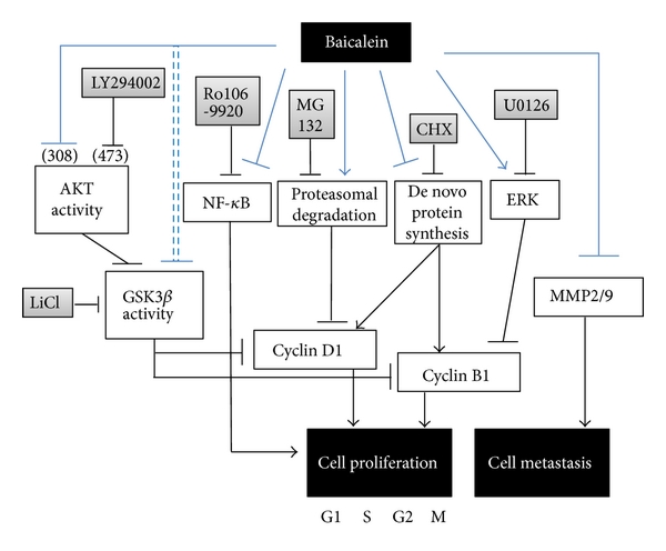
579751.fig.007fig7
Figure 7: The pathway scheme of baicalein on the cell proliferation inhibition and cell metastasis inhibition in human bladder cancer cell 5637. The anti-proliferation and anti-metastasis activities of baicalein may contribute the anti-bladder-tumor effect in vivo.
Abbreviations

FBS: Fetal bovine serum
MMP-2: Matrix metalloproteinase-2
MMP-9: Matrix metalloproteinase-9
MTT: 3-(4,5-dimethylthiazol-2-yl)2,5-diphenyltetrazolium bromide
NF-κB: Nuclear factor-kappaB
PI3K: Phosphatidylinositol 3-kinase.
Conflict of Interests

The authors declare that there is no conflict of interests.

Authors’ Contribution

J.-Y. Wu and K.-W. Tsai contributed equally to this work.

Acknowledgments

This work is supported by Grants from the National Science Council NSC101-2320-B-415-002-MY3 of the Republic of China and Buddhist Tzuchi Dalin General Hospital, Dalin Town, Chiayi, Taiwan.

References

1 A. Jemal, F. Bray, M. M. Center, J. Ferlay, E. Ward, and D. Forman, “Global cancer statistics,” CA Cancer Journal for Clinicians, vol. 61, no. 2, pp. 69–90, 2011. View at Publisher · View at Google Scholar · View at Scopus
2 R. Siegel, D. Naishadham, and A. Jemal, “Cancer statistics 2012,” A Cancer Journal for Clinicians, vol. 62, no. 1, pp. 10–29, 2012.
3 D. J. McConkey, S. Lee, W. Choi et al., “Molecular genetics of bladder cancer: emerging mechanisms of tumor initiation and progression,” Urologic Oncology, vol. 28, no. 4, pp. 429–440, 2010. View at Publisher· View at Google Scholar · View at Scopus
4 X. R. Wu, “Urothelial tumorigenesis: a tale of divergent pathways,” Nature Reviews Cancer, vol. 5, no. 9, pp. 713–725, 2005. View at Publisher · View at Google Scholar · View at Scopus
5 L. C. Pagliaro and P. Sharma, “Review of metastatic bladder cancer,” Minerva Urologica e Nefrologica, vol. 58, no. 1, pp. 53–71, 2006. View at Scopus
6 C. C. Lin and D. E. Shieh, “The anti-inflammatory activity of Scutellaria rivularis extracts and its active components, baicalin, baicalein and wogonin,” American Journal of Chinese Medicine, vol. 24, no. 1, pp. 31–36, 1996. View at Scopus
7 Z. H. Shao, T. L. Vanden Hoek, Y. Qin et al., “Baicalein attenuates oxidant stress in cardiomyocytes,” American Journal of Physiology, vol. 282, no. 3, pp. H999–H1006, 2002. View at Scopus
8 L. G. Chen, L. Y. Hung, K. W. Tsai et al., “Wogonin, a bioactive flavonoid in herbal tea, inhibits inflammatory cyclooxygenase-2 gene expression in human lung epithelial cancer cells,” Molecular Nutrition and Food Research, vol. 52, no. 11, pp. 1349–1357, 2008. View at Publisher · View at Google Scholar · View at Scopus
9 J. I. Chao, W. C. Su, and H. F. Liu, “Baicalein induces cancer cell death and proliferation retardation by the inhibition of CDC2 kinase and survivin associated with opposite role of p38 mitogen-activated protein kinase and AKT,” Molecular Cancer Therapeutics, vol. 6, no. 11, pp. 3039–3048, 2007. View at Publisher · View at Google Scholar · View at Scopus
10 R. H. Jiang, W. C. Su, H. F. Liu, H. S. Huang, and J. I. Chao, “Opposite expression of securin and γ-H2AX regulates baicalein-induced cancer cell death,” Journal of Cellular Biochemistry, vol. 111, no. 2, pp. 274–283, 2010. View at Publisher · View at Google Scholar · View at Scopus
11 H. Takahashi, M. C. Chen, H. Pham et al., “Baicalein, a component of Scutellaria baicalensis, induces apoptosis by Mcl-1 down-regulation in human pancreatic cancer cells,” Biochimica et Biophysica Acta, vol. 1813, no. 8, pp. 1465–1474, 2011. View at Publisher · View at Google Scholar · View at Scopus
12 Y. W. Chiu, T. H. Lin, W. S. Huang, et al., “Baicalein inhibits the migration and invasive properties of human hepatoma cells,” Toxicology and Applied Pharmacology, vol. 255, no. 3, pp. 316–326, 2011.
13 L. Wang, Y. Ling, Y. Chen et al., “Flavonoid baicalein suppresses adhesion, migration and invasion of MDA-MB-231 human breast cancer cells,” Cancer Letters, vol. 297, no. 1, pp. 42–48, 2010. View at Publisher · View at Google Scholar · View at Scopus
14 B. Wu, J. Li, D. Huang, et al., “Baicalein mediates inhibition of migration and invasiveness of skin carcinoma through Ezrin in A431 cells,” BMC Cancer, vol. 11, article 527, 2011.
15 J. T. Fox, S. Sakamuru, R. Huang, et al., “High-throughput genotoxicity assay identifies antioxidants as inducers of DNA damage response and cell death,” Proceedings of the National Academy of Sciences of the United States of America, vol. 109, no. 14, pp. 5423–5428, 2012.
16 J. Y. Wu, K. T. Chung, Y. W. Liu et al., “Synthesis and biological evaluation of novel C(6) modified baicalein derivatives as antioxidative agents,” Journal of Agricultural and Food Chemistry, vol. 56, no. 8, pp. 2838–2845, 2008. View at Publisher · View at Google Scholar · View at Scopus
17 C. H. Shen, J. J. Shee, J. Y. Wu, Y. W. Lin, J. D. Wu, and Y. W. Liu, “Combretastatin A-4 inhibits cell growth and metastasis in bladder cancer cells and retards tumour growth in a murine orthotopic bladder tumour model,” British Journal of Pharmacology, vol. 160, no. 8, pp. 2008–2027, 2010. View at Publisher · View at Google Scholar · View at Scopus
18 S. Y. Wu, Y. R. Lee, C. C. Huang, et al., “Curcumin-induced heme oxygenase-1 expression plays a negative role for its anti-cancer effect in bladder cancers,” Food and Chemical Toxicology, vol. 50, no. 10, pp. 3530–3536, 2012.
19 P. S. Klein and D. A. Melton, “A molecular mechanism for the effect of lithium on development,” Proceedings of the National Academy of Sciences of the United States of America, vol. 93, no. 16, pp. 8455–8459, 1996. View at Publisher · View at Google Scholar · View at Scopus
20 F. Zhan, C. J. Phiel, L. Spece, N. Gurvich, and P. S. Klein, “Inhibitory phosphorylation of glycogen synthase kinase-3 (GSK-3) in response to lithium: evidence for autoregulation of GSK-3,” Journal of Biological Chemistry, vol. 278, no. 35, pp. 33067–33077, 2003. View at Publisher · View at Google Scholar· View at Scopus
21 Q. M. Zhou, S. Wang, H. Zhang et al., “The combination of baicalin and baicalein enhances apoptosis via the ERK/p38 MAPK pathway in human breast cancer cells,” Acta Pharmacologica Sinica, vol. 30, no. 12, pp. 1648–1658, 2009. View at Publisher · View at Google Scholar · View at Scopus
22 Y. H. Cheng, L. A. Li, P. Lin, et al., “Baicalein induces G1 arrest in oral cancer cells by enhancing the degradation of cyclin D1 and activating AhR to decrease Rb phosphorylation,” Toxicology and Applied Pharmacology, vol. 263, no. 3, pp. 360–367, 2012.
23 H. W. C. Leung, W. H. Yang, M. Y. Lai, C. J. Lin, and H. Z. Lee, “Inhibition of 12-lipoxygenase during baicalein-induced human lung nonsmall carcinoma H460 cell apoptosis,” Food and Chemical Toxicology, vol. 45, no. 3, pp. 403–411, 2007. View at Publisher · View at Google Scholar · View at Scopus
24 B. C. Y. Wong, W. P. Wang, C. H. Cho et al., “12-Lipoxygenase inhibition induced apoptosis in human gastric cancer cells,” Carcinogenesis, vol. 22, no. 9, pp. 1349–1354, 2001. View at Scopus
25 M. Bonham, J. Posakony, I. Coleman, B. Montgomery, J. Simon, and P. S. Nelson, “Characterization of chemical constituents in Scutellaria baicalensis with antiandrogenic and growth-inhibitory activities toward prostate carcinoma,” Clinical Cancer Research, vol. 11, no. 10, pp. 3905–3914, 2005. View at Publisher · View at Google Scholar · View at Scopus
26 S. Chen, Q. Ruan, E. Bedner et al., “Effects of the flavonoid baicalin and its metabolite baicalein on androgen receptor expression, cell cycle progression and apoptosis of prostate cancer cell lines,” Cell Proliferation, vol. 34, no. 5, pp. 293–304, 2001. View at Publisher · View at Google Scholar · View at Scopus
27 M. K. Roy, K. Nakahara, V. Na Thalang et al., “Baicalein, a flavonoid extracted from a methanolic extract of Oroxylum indicum inhibits proliferation of a cancer cell line in vitro via induction of apoptosis,” Pharmazie, vol. 62, no. 2, pp. 149–153, 2007. View at Publisher · View at Google Scholar · View at Scopus
28 W. H. Chang, C. H. Chen, and F. J. Lu, “Different effects of baicalein, baicalin and wogonin on mitochondrial function, glutathione content and cell cycle progression in human hepatoma cell lines,” Planta Medica, vol. 68, no. 2, pp. 128–132, 2002. View at Publisher · View at Google Scholar · View at Scopus
29 Z. Ma, K. I. Otsuyama, S. Liu et al., “Baicalein, a component of Scutellaria radix from Huang-Lian-Jie-Du-Tang (HLJDT), leads to suppression of proliferation and induction of apoptosis in human myeloma cells,” Blood, vol. 105, no. 8, pp. 3312–3318, 2005. View at Publisher · View at Google Scholar · View at Scopus
30 M. Himeji, T. Ohtsuki, H. Fukazawa et al., “Difference of growth-inhibitory effect of Scutellaria baicalensis-producing flavonoid wogonin among human cancer cells and normal diploid cell,” Cancer Letters, vol. 245, no. 1-2, pp. 269–274, 2007. View at Publisher · View at Google Scholar · View at Scopus
31 D. Kong, Y. Zhang, T. Yamori, H. Duan, and M. Jin, “Inhibitory activity of flavonoids against class I phosphatidylinositol 3-kinase isoforms,” Molecules, vol. 16, no. 6, pp. 5159–5167, 2011. View at Publisher· View at Google Scholar · View at Scopus
32 G. P. Pidgeon, M. Kandouz, A. Meram, and K. V. Honn, “Mechanisms controlling cell cycle arrest and induction of apoptosis after 12-lipoxygenase inhibition in prostate cancer cells,” Cancer Research, vol. 62, no. 9, pp. 2721–2727, 2002. View at Scopus
33 L. Stephens, K. Anderson, D. Stokoe et al., “Prohtein kinase B kinases that mediate phosphatidylinositol 3,4,5-trisphosphate-dependent activation of protein kinase B,” Science, vol. 279, no. 5351, pp. 710–714, 1998. View at Publisher · View at Google Scholar · View at Scopus
34 D. D. Sarbassov, D. A. Guertin, S. M. Ali, and D. M. Sabatini, “Phosphorylation and regulation of Akt/PKB by the rictor-mTOR complex,” Science, vol. 307, no. 5712, pp. 1098–1101, 2005. View at Publisher · View at Google Scholar · View at Scopus
35 R. S. Jope, C. J. Yuskaitis, and E. Beurel, “Glycogen synthase kinase-3 (GSK3): inflammation, diseases, and therapeutics,” Neurochemical Research, vol. 32, no. 4-5, pp. 577–595, 2007. View at Publisher · View at Google Scholar · View at Scopus
36 C. J. Hsieh, K. Hall, T. Ha, C. Li, G. Krishnaswamy, and D. S. Chi, “Baicalein inhibits IL-1β- and TNF-α-induced inflammatory cytokine production from human mast cells via regulation of the NF-κB pathway,” Clinical and Molecular Allergy, vol. 5, article 5, 2007. View at Publisher · View at Google Scholar · View at Scopus
37 B. Y. Kang, S. W. Chung, S. H. Kim, D. Cho, and T. S. Kim, “Involvement of nuclear factor-κB in the inhibition of interleukin-12 production from mouse macrophages by baicalein, a flavonoid in Scutellaria baicalensis,” Planta Medica, vol. 69, no. 8, pp. 687–691, 2003. View at Publisher · View at Google Scholar · View at Scopus
38 M. H. Kim, S. Y. Ryu, M. A. Bae, J. S. Choi, Y. K. Min, and S. H. Kim, “Baicalein inhibits osteoclast differentiation and induces mature osteoclast apoptosis,” Food and Chemical Toxicology, vol. 46, no. 11, pp. 3375–3382, 2008. View at Publisher · View at Google Scholar · View at Scopus
39 C. J. Chen, S. L. Raung, S. L. Liao, and S. Y. Chen, “Inhibition of inducible nitric oxide synthase expression by baicalein in endotoxin/cytokine-stimulated microglia,” Biochemical Pharmacology, vol. 67, no. 5, pp. 957–965, 2004. View at Publisher · View at Google Scholar · View at Scopus
40 K. Suk, H. Lee, S. S. Kang, G. J. Cho, and W. S. Choi, “Flavonoid baicalein attenuates activation-induced cell death of brain microglia,” Journal of Pharmacology and Experimental Therapeutics, vol. 305, no. 2, pp. 638–645, 2003. View at Publisher · View at Google Scholar · View at Scopus
41 J. I. Inoue, J. Gohda, T. Akiyama, and K. Semba, “NF-κB activation in development and progression of cancer,” Cancer Science, vol. 98, no. 3, pp. 268–274, 2007. View at Publisher · View at Google Scholar · View at Scopus
42 G. Sethi, B. Sung, and B. B. Aggarwal, “Nuclear factor-κB activation: from bench to bedside,” Experimental Biology and Medicine, vol. 233, no. 1, pp. 21–31, 2008. View at Publisher · View at Google Scholar · View at Scopus
43 J. Deng, W. Xia, S. A. Miller, Y. Wen, H. Y. Wang, and M. C. Hung, “Crossregulation of NF-κB by the APC/GSK-3β/β-catenin pathway,” Molecular Carcinogenesis, vol. 39, no. 3, pp. 139–146, 2004. View at Publisher · View at Google Scholar · View at Scopus
44 H. Z. Lee, H. W. C. Leung, M. Y. Lai, and C. H. Wu, “Baicalein induced cell cycle arrest and apoptosis in human lung squamous carcinoma CH27 cells,” Anticancer Research, vol. 25, no. 2, pp. 959–964, 2005.View at Scopus
45 C. S. Ong, J. Zhou, C. N. Ong, and H. M. Shen, “Luteolin induces G1 arrest in human nasopharyngeal carcinoma cells via the Akt-GSK-3β-cyclin D1 pathway,” Cancer Letters, vol. 298, no. 2, pp. 167–175, 2010. View at Publisher · View at Google Scholar · View at Scopus

Sanguinarine (See also chelerythrine)

Cancer:
Prostate, bladder, breast, colon, melanoma, leukemia

Action: Pro-oxidative, anti-inflammatory, apoptosis induction

AR+/AR- Prostate Cancer

Sanguinarine, a benzophenanthridine alkaloid derived from the bloodroot plant Sanguinaria canadensis (L.), has been shown to possess anti-microbial, anti-inflammatory, anti-cancer and anti-oxidant properties. It has been shown that sanguinarine possesses strong anti-proliferative and pro-apoptotic properties against human epidermoid carcinoma A431 cells and immortalized human HaCaT keratinocytes. Employing androgen-responsive human prostate carcinoma LNCaP cells and androgen-unresponsive human prostate carcinoma DU145 cells, the anti-proliferative properties of sanguinarine against prostate cancer were also examined.

The mechanism of the anti-proliferative effects of sanguinarine against prostate cancer were examined by determining the effect of sanguinarine on critical molecular events known to regulate the cell-cycle and the apoptotic machinery.

A highlight of this study was the fact that sanguinarine induced growth-inhibitory and anti-proliferative effects in human prostate carcinoma cells irrespective of their androgen status. To our knowledge, this is the first study showing the involvement of cyclin kinase inhibitor-cyclin-cyclin-dependent kinase machinery during cell-cycle arrest and apoptosis of prostate cancer cells by sanguinarine. These results suggest that sanguinarine may be developed as an agent for the management of prostate cancer (Adhami et al., 2004).

Breast Cancer

The effects of this compound were examined on reactive oxygen species (ROS) production and its association with apoptotic tumor cell death using a human breast carcinoma MDA-MB-231 cell line. Cytotoxicity was evaluated by trypan blue exclusion methods. Apoptosis was detected using DAPI staining, agarose gel electrophoresis and flow cytometer. The expression levels of proteins were determined by Western blot analyzes and caspase activities were measured using colorimetric assays.

These observations clearly indicate that ROS is involved in the early molecular events in the sanguinarine-induced apoptotic pathway. Data suggests that sanguinarine-induced ROS are key mediators of MMP collapse, which leads to the release of cytochrome c followed by caspase activation, culminating in apoptosis (Choi, Kim, Lee & Choi, 2008).

Leukemia

Sanguinarine, chelerythrine and chelidonine are isoquinoline alkaloids derived from the greater celandine. They possess a broad spectrum of pharmacological activities. It has been shown that their anti-tumor activity is mediated via different mechanisms, which can be promising targets for anti-cancer therapy.

This study focuses on the differential effects of these alkaloids upon cell viability, DNA damage, and nucleus integrity in mouse primary spleen and lymphocytic leukemic cells, L1210. Sanguinarine and chelerythrine produced a dose-dependent increase in DNA damage and cytotoxicity in both primary mouse spleen cells and L1210 cells. Chelidonine did not show a significant cytotoxicity or damage DNA in both cell types, but completely arrested growth of L1210 cells.

Data suggests that cytotoxic and DNA-damaging effects of chelerythrine and sanguinarine are more selective against mouse leukemic cells and primary mouse spleen cells, whereas chelidonine blocks proliferation of L1210 cells. The action of chelidonine on normal and tumor cells requires further investigation (Kaminsky, Lin, Filyak, & Stoika, 2008).

T-lymphoblastic Leukemia

Apoptogenic and DNA-damaging effects of chelidonine (CHE) and sanguinarine (SAN), two structurally related benzophenanthridine alkaloids isolated from Chelidonium majus, were compared. Both alkaloids induced apoptosis in human acute T-lymphoblastic leukemia MT-4 cells. Apoptosis induction by CHE and SAN in these cells were accompanied by caspase-9 and -3 activation and an increase in the pro-apoptotic Bax protein. An elevation in the percentage of MT-4 cells possessing caspase-3 in active form after their treatment with CHE or SAN was in parallel to a corresponding increase in the fraction of apoptotic cells.

The involvement of the mitochondria in apoptosis induction by both alkaloids was supported by cytochrome C elevation in cytosol, with an accompanying decrease in cytochrome C content in the mitochondrial fraction. At the same time, two alkaloids under study differed drastically in their cell-cycle phase-specific effects, since only CHE arrested MT-4 cells at the G2/M phase. It was previously demonstrated, that CHE, in contrast to SAN, does not interact directly with DNA. (Philchenkov, Kaminskyy, Zavelevich, & Stoika, 2008).

Sanguinarine, chelerythrine and chelidonine possess prominent apoptotic effects towards cancer cells. This study found that sanguinarine and chelerythrine induced apoptosis in human CEM T-leukemia cells, accompanied by an early increase in cytosolic cytochrome C that precedes caspases-8, -9 and -3 processing. Effects of sanguinarine and chelerythrine on mitochondria were confirmed by clear changes in morphology (3h), however chelidonine did not affect mitochondrial integrity.

Sanguinarine and chelerythrine also caused marked DNA damage in cells after 1h, but a more significant increase in impaired cells occurred after 6h. Chelidonine induced intensive DNA damage in 15–20% cells after 24h. Results demonstrated that rapid cytochrome C release in CEM T-leukemia cells exposed to sanguinarine or chelerythrine was not accompanied by changes in Bax, Bcl-2 and Bcl-X((L/S)) proteins in the mitochondrial fraction, and preceded activation of the initiator caspase-8 (Kaminskyy, Kulachkovskyy & Stoika, 2008).

Colorectal Cancer

The effects of sanguinarine, a benzophenanthridine alkaloid, was examined on reactive oxygen species (ROS) production, and the association of these effects with apoptotic cell death, in a human colorectal cancer HCT-116 cell line. Sanguinarine generated ROS, followed by a decrease in mitochondrial membrane potential (MMP), activation of caspase-9 and -3, and down-regulation of anti-apoptotic proteins, such as Bcl2, XIAP and cIAP-1. Sanguinarine also promoted the activation of caspase-8 and truncation of Bid (tBid).

Observations clearly indicate that ROS, which are key mediators of Egr-1 activation and MMP collapse, are involved in the early molecular events in the sanguinarine-induced apoptotic pathway acting in HCT-116 cells (Han, Kim, Yoo, & Choi, 2013).

Bladder Cancer

Although the effects of sanguinarine, a benzophenanthridine alkaloid, on the inhibition of some kinds of cancer cell growth have been established, the underlying mechanisms are not completely understood. This study investigated possible mechanisms by which sanguinarine exerts its anti-cancer action in cultured human bladder cancer cell lines (T24, EJ, and 5637). Sanguinarine treatment resulted in concentration-response growth inhibition of the bladder cancer cells by inducing apoptosis.

Taken together, the data provide evidence that sanguinarine is a potent anti-cancer agent, which inhibits the growth of bladder cancer cells and induces their apoptosis through the generation of free radicals (Han et al., 2013).

Melanoma

Sanguinarine is a natural isoquinoline alkaloid derived from the root of Sanguinaria canadensis and from other poppy fumaria species, and is known to have a broad spectrum of pharmacological properties. Current study has found that sanguinarine, at low micromolar concentrations, showed a remarkably rapid killing activity against human melanoma cells. Sanguinarine disrupted the mitochondrial transmembrane potential (ΔΨ m), released cytochrome C and Smac/DIABLO from mitochondria to cytosol, and induced oxidative stress. Thus, pre-treatment with the thiol anti-oxidants NAC and GSH abrogated the killing activity of sanguinarine. Collectively, data suggests that sanguinarine is a very rapid inducer of human melanoma caspase-dependent cell death that is mediated by oxidative stress (Burgeiro, Bento, Gajate, Oliveira, & Mollinedo, 2013).

References

Adhami YM, Aziz MH, Reagan-Shaw SR, et al. (2004). Sanguinarine causes cell-cycle blockade and apoptosis of human prostate carcinoma cells via modulation of cyclin kinase inhibitor-cyclin-cyclin-dependent kinase machinery. Mol Cancer Ther, 3:933


Burgeiro A, Bento AC, Gajate C, Oliveira PJ, Mollinedo F. (2013). Rapid human melanoma cell death induced by sanguinarine through oxidative stress. European Journal of Pharmacology, 705(1-3), 109-18. doi: 10.1016/j.ejphar.2013.02.035.


Choi WY, Kim GY, Lee WH, Choi YH. (2008). Sanguinarine, a benzophenanthridine alkaloid, induces apoptosis in MDA-MB-231 human breast carcinoma cells through a reactive oxygen species-mediated mitochondrial pathway. Chemotherapy, 54(4), 279-87. doi: 10.1159/000149719.


Han MH, Kim GY, Yoo YH, Choi YH. (2013). Sanguinarine induces apoptosis in human colorectal cancer HCT-116 cells through ROS-mediated Egr-1 activation and mitochondrial dysfunction. Toxicology Letters, 220(2), 157-66. doi: 10.1016/j.toxlet.2013.04.020.


Han MH, Park C, Jin CY, et al. (2013). Apoptosis induction of human bladder cancer cells by sanguinarine through reactive oxygen species-mediated up-regulation of early growth response gene-1. PLoS One, 8(5), e63425. doi: 10.1371/journal.pone.0063425.


Kaminskyy V, Lin KW, Filyak Y, Stoika R. (2008). Differential effect of sanguinarine, chelerythrine and chelidonine on DNA damage and cell viability in primary mouse spleen cells and mouse leukemic cells. Cell Biology International, 32(2), 271-277.


Kaminskyy V, Kulachkovskyy O, Stoika R. (2008) A decisive role of mitochondria in defining rate and intensity of apoptosis induction by different alkaloids. Toxicology Letters, 177(3), 168-81. doi: 10.1016/j.toxlet.2008.01.009.


Philchenkov A, Kaminskyy V, Zavelevich M, Stoika R. (2008). Apoptogenic activity of two benzophenanthridine alkaloids from Chelidonium majus L. does not correlate with their DNA-damaging effects. Toxicology In Vitro, 22(2), 287-95.

Matricaria chamomilla/Matricaria recutita

Cancer: Colorectal., ovarian, testicular, bladder, lung

Action: Neuropathy, anti-inflammatory

Colorectal Cancer; Ovarian Cancer; Testicular Cancer; Bladder Cancer; Lung Cancer; Chemotherapy

Studies have shown that cisplatin could have painful effects on human and animal models. Matricaria chamomilla (MC) has analgesic and anti-inflammatory effects, and may hence be an effective treatment for ciplatin-induced peripheral neuropathy as a replacement for morphine. Experiments were performed on 60 NMRI male mice weighed 25 g to 30 g, which have been divided into 6 groups. The first group received normal saline; the second group received MC hydroalcoholic extract; the third group received cisplatin; the fourth group received MC hydroalcoholic extract and cisplatin, 96 hours before formalin test; the fifth group received morphine and the sixth group received cisplatin and morphine.

Results showed that formalin induced significant (P < 0.05) pain response (the first phase: 0–5 min and the second phase: 15–40 min after injection). Administration of MC extract before formalin injection showed significant (P < 0.05) decrease of pain responses in the first and second phase. Administration of cisplatin produced significant (P < 0.05) increase in pain response in both phases of formalin test. Injection of MC extract and cisplatin together have shown that MC is able to decrease the second phase of cisplatin-induced pain significantly (P < 0.05).

In comparison morphine has analgesic effects in the first phase and MC extract has anti- inflammatory effects in the second phase of formalin test significantly (P < 0.05). MC and cisplatin have analgesic and painful neuropathic respective effects, and MC hydroalcoholic extract is able to decrease cisplatin-induced pain and inflammation better than morphine (Abad et al., 2011).

Anti-inflammatory

Flavonoid-7-glycosides, major constituents of chamomile flowers, may be responsible for the anti-inflammatory action, which is due to the inhibition of neutrophil elastase and gastric metalloproteinase-9 activity and secretion; the inhibition occurring in a concentration dependent manner (Bulgari et al., 2012).

The anti-cancer properties of aqueous and methanolic extracts of Matricaria chamomilla against various human cancer cell lines were investigated. Chamomile exposure resulted in differential apoptosis in cancer cells but not in normal cells at similar doses. HPLC analysis of chamomile extract confirmed apigenin 7-O-glucoside as the major constituent of chamomile; some minor glycoside components were also observed. Apigenin glucosides inhibited cancer cell growth but to a lesser extent than the parent aglycone, apigenin (Srivastava & Gupta, 2007).

References

Abad NA, Nouri MHK, Gharjanie A, Tavakoli F. (2011). Effect of Matricaria chamomilla Hydroalcoholic Extract on Cisplatin-induced Neuropathy in Mice. Chinese Journal of Natural Medicines, 9(2):126–131


Bulgari M, Sangiovanni E, Colombo E, et al. (2012). Inhibition of neutrophil elastase and metalloprotease-9 of human adenocarcinoma gastric cells by chamomile (Matricaria recutita L.) infusion. Phytother Res, 26(12):1817-22. doi: 10.1002/ptr.4657.


Srivastava JK, Gupta S. (2007). Anti-proliferative and apoptotic effects of chamomile extract in various human cancer cells. J Agric Food Chem, 55(23):9470-8.

Magnolol

Cancer:
Bladder, breast, colon, prostate, glioblastoma, ovarian, leukemia, lung

Action: Anti-inflammatory, apoptosis, inhibits angiogenesis, anti-metastatic

Magnolol (Mag), an active constituent isolated from the Chinese herb hou po (Magnolia officinalis (Rehder & Wilson)) has long been used to suppress inflammatory processes. It has anti-cancer activity in colon, hepatoma, and leukemia cell lines.

Anti-inflammatory

Magnolol (Mag) suppressed IL-6-induced promoter activity of cyclin D1 and monocyte chemotactic protein (MCP)-1 for which STAT3 activation plays a role. Pre-treatment of ECs with Mag dose-dependently inhibited IL-6-induced Tyr705 and Ser727 phosphorylation in STAT3 without affecting the phosphorylation of JAK1, JAK2, and ERK1/2. Mag pre-treatment of these ECs dose-dependently suppressed IL-6-induced promoter activity of intracellular cell adhesion molecule (ICAM)-1 that contains functional IL-6 response elements (IREs).

In conclusion, our results indicate that Mag inhibits IL-6-induced STAT3 activation and subsequently results in the suppression of downstream target gene expression in ECs. These results provide a therapeutic basis for the development of Mag as an anti-inflammatory agent for vascular disorders including atherosclerosis (Chen et al., 2006).

Bladder Cancer; Inhibits Angiogenesis

In the present study, Chen et al. (2013) demonstrated that magnolol significantly inhibited angiogenesis in vitro and in vivo, evidenced by the attenuation of hypoxia and vascular endothelial growth factor (VEGF)-induced tube formation of human umbilical vascular endothelial cells, vasculature generation in chicken chorioallantoic membrane, and Matrigel plug.

In hypoxic human bladder cancer cells (T24), treatment with magnolol inhibited hypoxia-stimulated H2O2 formation, HIF-1α induction including mRNA, protein expression, and transcriptional activity as well as VEGF secretion. Interestingly, magnolol also acts as a VEGFR2 antagonist, and subsequently attenuates the downstream AKT/mTOR/p70S6K/4E-BP-1 kinase activation both in hypoxic T24 cells and tumor tissues. As expected, administration of magnolol greatly attenuated tumor growth, angiogenesis and the protein expression of HIF-1α, VEGF, CD31, a marker of endothelial cells, and carbonic anhydrase IX, an endogenous marker for hypoxia, in the T24 xenograft mouse model.

Collectively, these findings strongly indicate that the anti-angiogenic activity of magnolol is, at least in part, mediated by suppressing HIF-1α/VEGF-dependent pathways, and suggest that magnolol may be a potential drug for human bladder cancer therapy.

Colon Cancer; Induces Apoptosis

Emerging evidence has suggested that activation of AMP-activated protein kinase (AMPK), a potential cancer therapeutic target, is involved in apoptosis in colon cancer cells. However, the effects of magnolol on human colon cancer through activation of AMPK remain unexplored.

Magnolol displayed several apoptotic features, including propidium iodide labeling, DNA fragmentation, and caspase-3 and poly(ADP-ribose) polymerase cleavages. Park et al. (2012) showed that magnolol induced the phosphorylation of AMPK in dose- and time-dependent manners.

Magnolol down-regulated expression of the anti-apoptotic protein Bcl2, up-regulated expression of pro-apoptotic protein p53 and Bax, and caused the release of mitochondrial cytochrome c. Magnolol-induced p53 and Bcl2 expression was abolished in the presence of compound C. Magnolol inhibited migration and invasion of HCT-116 cells through AMPK activation. These findings demonstrate that AMPK mediates the anti-cancer effects of magnolol through apoptosis in HCT-116 cells.

Ovarian Cancer

Treatment of HER-2 overexpressing ovarian cancer cells with magnolol down-regulated the HER-2 downstream PI3K/Akt signaling pathway, and suppressed the expression of downstream target genes, vascular endothelial growth factor (VEGF), matrix metalloproteinase 2 (MMP2) and cyclin D1. Consistently, magnolol-mediated inhibition of MMP2 activity could be prevented by co-treatment with epidermal growth factor. Migration assays revealed that magnolol treatment markedly reduced the motility of HER-2 overexpressing ovarian cancer cells. These findings suggest that magnolol may act against HER-2 and its downstream PI3K/Akt/mTOR-signaling network, thus resulting in suppression of HER-2mediated transformation and metastatic potential in HER-2 overexpressing ovarian cancers. These results provide a novel mechanism to explain the anti-cancer effect of magnolol (Chuang et al., 2011).

Lung Cancer

Magnolol has been found to inhibit cell growth, increase lactate dehydrogenase release, and modulate cell cycle in human lung carcinoma A549 cells. Magnolol induced the activation of caspase-3 and cleavage of Poly-(ADP)-ribose polymerase, and decreased the expression level of nuclear factor-κB/Rel A in the nucleus. In addition, magnolol inhibited basic fibroblast growth factor-induced proliferation and capillary tube formation of human umbilical vein endothelial cells. These data indicate that magnolol is a potential candidate for the treatment of human lung carcinoma (Seo et al., 2011).

Prostate Cancer; Anti-metastatic

Matrix metalloproteinases (MMPs) are enzymes involved in various steps of metastasis development. The objective of this study was to study the effects of magnolol on cancer invasion and metastasis using PC-3 human prostate carcinoma cells. Magnolol inhibited cell growth in a dose-dependent manner. In an invasion assay conducted in Transwell chambers, magnolol showed 33 and 98% inhibition of cancer cell at 10 microM and 20 microM concentrations, respectively, compared to the control. The protein and mRNA levels of both MMP-2 and MMP-9 were down-regulated by magnolol treatment in a dose-dependent manner.

These results demonstrate the anti-metastatic properties of magnolol in inhibiting the adhesion, invasion, and migration of PC-3 human prostate cancer cells (Hwang et al., 2010).

Glioblastoma Cancer

Magnolol has been found to concentration-dependently (0-40 microM) decrease the cell number in a cultured human glioblastoma cancer cell line (U373) and arrest the cells at the G0/G1 phase of the cell-cycle.

Pre-treatment of U373 with p21/Cip1 specific antisense oligodeoxynucleotide prevented the magnolol-induced increase of p21/Cip1 protein levels and the decrease of DNA synthesis. Magnolol at a concentration of 100 microM induced DNA fragmentation in U373. These findings suggest the potential applications of magnolol in the treatment of human brain cancers (Chen et al. 2011).

Inhibits Angiogenesis

Magnolol inhibited VEGF-induced Ras activation and subsequently suppressed extracellular signal-regulated kinase (ERK), phosphatidylinositol-3-kinase (PI3K)/Akt and p38, but not Src and focal adhesion kinase (FAK). Interestingly, the knockdown of Ras by short interfering RNA produced inhibitory effects that were similar to the effects of magnolol on VEGF-induced angiogenic signaling events, such as ERK and Akt/eNOS activation, and resulted in the inhibition of proliferation, migration, and vessel sprouting in HUVECs.

In combination, these results demonstrate that magnolol is an inhibitor of angiogenesis and suggest that this compound could be a potential candidate in the treatment of angiogenesis-related diseases (Kim et al., 2013).

References

Chen LC, Liu YC, Liang YC, Ho YS, Lee WS. (2009). Magnolol inhibits human glioblastoma cell proliferation through up-regulation of p21/Cip1. J Agric Food Chem, 57(16):7331-7. doi: 10.1021/jf901477g.


Chen MC, Lee CF, Huang WH, Chou TC. (2013). Magnolol suppresses hypoxia-induced angiogenesis via inhibition of HIF-1 α /VEGF signaling pathway in human bladder cancer cells. Biochem Pharmacol, 85(9):1278-87. doi: 10.1016/j.bcp.2013.02.009.


Chen SC, Chang YL, Wang DL, Cheng JJ. (2006). Herbal remedy magnolol suppresses IL-6-induced STAT3 activation and gene expression in endothelial cells. Br J Pharmacol, 148(2): 226–232. doi: 10.1038/sj.bjp.0706647


Chuang TC, Hsu SC, Cheng YT, et al. (2011). Magnolol down-regulates HER2 gene expression, leading to inhibition of HER2-mediated metastatic potential in ovarian cancer cells. Cancer Lett, 311(1):11-9. doi: 10.1016/j.canlet.2011.06.007.


Hwang ES, Park KK. (2010). Magnolol suppresses metastasis via inhibition of invasion, migration, and matrix metalloproteinase-2/-9 activities in PC-3 human prostate carcinoma cells. Biosci Biotechnol Biochem, 74(5):961-7.


Kim KM, Kim NS, Kim J, et al. (2013). Magnolol Suppresses Vascular Endothelial Growth Factor-Induced Angiogenesis by Inhibiting Ras-Dependent Mitogen-Activated Protein Kinase and Phosphatidylinositol 3-Kinase/Akt Signaling Pathways. Nutr Cancer.


Park JB, Lee MS, Cha EY, et al. (2012). Magnolol-induced apoptosis in HCT-116 colon cancer cells is associated with the AMP-activated protein kinase signaling pathway. Biol Pharm Bull, 35(9):1614-20.


Seo JU, Kim MH, Kim HM, Jeong HJ. (2011). Anti-cancer potential of magnolol for lung cancer treatment. Arch Pharm Res, 34(4):625-33. doi: 10.1007/s12272-011-0413-8.

Hispolon

Cancer: Bladder, breast, liver, gastric

Action: Anti-inflammatory, cytostatic, cytotoxic, pro-oxidative, anti-proliferative

Hispolon is an active phenolic compound of Phellinus igniarius , a mushroom that has recently been shown to have anti-oxidant, anti-inflammatory, and anti-cancer activities.

Liver Cancer

Hispolon inhibited cellular growth of Hep3B cells in a time-dependent and dose-dependent manner, through the induction of cell-cycle arrest at S phase measured using flow cytometric analysis and apoptotic cell death, as demonstrated by DNA laddering. Exposure of Hep3B cells to hispolon resulted in apoptosis as evidenced by caspase activation, PARP cleavage, and DNA fragmentation. Hispolon treatment also activated JNK, p38 MAPK, and ERK expression. Inhibitors of ERK (PB98095), but not those of JNK (SP600125) and p38 MAPK (SB203580), suppressed hispolon-induced S-phase arrest and apoptosis in Hep3B cells.

These findings establish a mechanistic link between the MAPK pathway and hispolon-induced cell-cycle arrest and apoptosis in Hep3B cells (Huang et al., 2011).

Gastric Cancer, Breast Cancer, Bladder Cancer

Hispolon extracted from Phellinus species was found to induce epidermoid and gastric cancer cell apoptosis. Hispolon has also been found to inhibit breast and bladder cancer cell growth, regardless of p53 status. Furthermore, p21(WAF1), a cyclin-dependent kinase inhibitor, was elevated in hispolon-treated cells. MDM2, a negative regulator of p21(WAF1), was ubiquitinated and degraded after hispolon treatment.

Lu et al. (2009) also found that activated ERK1/2 (extracellular signal-regulated kinase1/2) was recruited to MDM2 and involved in mediating MDM2 ubiquitination. The results indicated that cells with higher ERK1/2 activity were more sensitive to hispolon. In addition, hispolon-induced caspase-7 cleavage was inhibited by the ERK1/2 inhibitor, U0126.

In conclusion, hispolon ubiquitinates and down-regulates MDM2 via MDM2-recruited activated ERK1/2. Therefore, hispolon may be a potential anti-tumor agent in breast and bladder cancers.

Gastric Cancer

The efficacy of hispolon in human gastric cancer cells and cell death mechanism was explored. Hispolon induced ROS-mediated apoptosis in gastric cancer cells and was more toxic toward gastric cancer cells than toward normal gastric cells, suggesting greater susceptibility of the malignant cells.

The mechanism of hispolon-induced apoptosis was that hispolon abrogated the glutathione anti-oxidant system and caused massive ROS accumulation in gastric cancer cells. Excessive ROS caused oxidative damage to the mitochondrial membranes and impaired the membrane integrity, leading to cytochrome c release, caspase activation, and apoptosis. Furthermore, hispolon potentiated the cytotoxicity of chemotherapeutic agents used in the clinical management of gastric cancer.

These results suggest that hispolon could be useful for the treatment of gastric cancer either as a single agent or in combination with other anti-cancer agents (Chen et al., 2008).

Anti-proliferative Activity

Hispolon, which lacks one aromatic unit in relation to curcumin, exhibits enhanced anti-inflammatory and anti-proliferative activities. Dehydroxy hispolon was least potent for all three activities. Overall the results indicate that the substitution of a hydroxyl group for a methoxy group at the meta positions of the phenyl rings in curcumin significantly enhanced the anti-inflammatory activity, and the removal of phenyl ring at the 7(th) position of the heptadiene back bone and addition of hydroxyl group significantly increased the anti-proliferative activity of curcumin and hispolon (Ravindran et al., 2010).

References

Chen W, Zhao Z, Li L, et al. (2008). Hispolon induces apoptosis in human gastric cancer cells through a ROS-mediated mitochondrial pathway. Free Radic Biol Med, 45(1):60-72. doi: 10.1016/j.freeradbiomed.2008.03.013.


Huang GJ, Deng JS, Huang SS, Hu ML. (2011). Hispolon induces apoptosis and cell-cycle arrest of human hepatocellular carcinoma Hep3B cells by modulating ERK phosphorylation. J Agric Food Chem, 59(13):7104-13. doi: 10.1021/jf201289e.


Lu TL, Huang GJ, Lu TJ, et al. (2009). Hispolon from Phellinus linteus has anti-proliferative effects via MDM2-recruited ERK1/2 activity in breast and bladder cancer cells. Food Chem Toxicol, 47(8):2013-21. doi: 10.1016/j.fct.2009.05.023.


Ravindran J, Subbaraju GV, Ramani MV, et al. (2010). Bisdemethylcurcumin and structurally related hispolon analogues of curcumin exhibit enhanced prooxidant, anti-proliferative and anti-inflammatory activities in vitro. Biochem Pharmacol, 79(11):1658-66. doi: 10.1016/j.bcp.2010.01.033.

Dietary Flavones

Cancer:
Prostate, colorectal., breast, pancreatic, bladder, ovarian, leukemia, liver, glioma, osteosarcoma, melanoma

Action: Anti-inflammatory, TAM resistance, cancer stem cells, down-regulate COX-2, apoptosis, cell-cycle arrest, anti-angiogenic, chemo-sensitzer, adramycin (ADM) resistance

Sulforaphane, Phenethyl isothiocyanate (PEITC), quercetin, epicatechin, catechin, Luteolin, apigenin

Anti-inflammatory

The anti-inflammatory activities of celery extracts, some rich in flavone aglycones and others rich in flavone glycosides, were tested on the inflammatory mediators tumor necrosis factor α (TNF-α) and nuclear factor kappa-light-chain-enhancer of activated B cells (NF-κB) in lipopolysaccharide-stimulated macrophages. Pure flavone aglycones and aglycone-rich extracts effectively reduced TNF-α production and inhibited the transcriptional activity of NF-κB, while glycoside-rich extracts showed no significant effects.

Celery diets with different glycoside or aglycone contents were formulated and absorption was evaluated in mice fed with 5% or 10% celery diets. Relative absorption in vivo was significantly higher in mice fed with aglycone-rich diets as determined by HPLC-MS/MS (where MS/MS is tandem mass spectrometry). These results demonstrate that deglycosylation increases absorption of dietary flavones in vivo and modulates inflammation by reducing TNF-α and NF-κB, suggesting the potential use of functional foods rich in flavones for the treatment and prevention of inflammatory diseases (Hostetler et al., 2012).

Colorectal Cancer

Association between the 6 main classes of flavonoids and the risk of colorectal cancer was examined using data from a national prospective case-control study in Scotland, including 1,456 incident cases and 1,456 population-based controls matched on age, sex, and residence area.

Dietary, including flavonoid, data were obtained from a validated, self-administered food frequency questionnaire. Risk of colorectal cancer was estimated using conditional logistic regression models in the whole sample and stratified by sex, smoking status, and cancer site and adjusted for established and putative risk factors.

The significant dose-dependent reductions in colorectal cancer risk that were associated with increased consumption of the flavonols quercetin, catechin, and epicatechin, remained robust after controlling for overall fruit and vegetable consumption or for other flavonoid intake. The risk reductions were greater among nonsmokers, but no interaction beyond a multiplicative effect was present.

This was the first of several a priori hypotheses to be tested in this large study and showed strong and linear inverse associations of flavonoids with colorectal cancer risk (Theodoratou et al., 2007).

Anti-angiogenic, Prostate Cancer

Luteolin is a common dietary flavonoid found in fruits and vegetables. The anti-angiogenic activity of luteolin was examined using in vitro, ex vivo, and in vivo models. Angiogenesis, the formation of new blood vessels from pre-existing vascular beds, is essential for tumor growth, invasion, and metastasis; hence, examination of this mechanism of tumor growth is essential to understanding new chemo-preventive targets. In vitro studies using rat aortic ring assay showed that luteolin at non-toxic concentrations significantly inhibited microvessel sprouting and proliferation, migration, invasion and tube formation of endothelial cells, which are key events in the process of angiogenesis. Luteolin also inhibited ex vivo angiogenesis as revealed by chicken egg chorioallantoic membrane assay (CAM) and matrigel plug assay.

Pro-inflammatory cytokines such as IL-1β, IL-6, IL-8, and TNF-α level were significantly reduced by the treatment of luteolin in PC-3 cells. Luteolin (10 mg/kg/d) significantly reduced the volume and the weight of solid tumors in prostate xenograft mouse model, indicating that luteolin inhibited tumorigenesis by targeting angiogenesis. Moreover, luteolin reduced cell viability and induced apoptosis in prostate cancer cells, which were correlated with the down-regulation of AKT, ERK, mTOR, P70S6K, MMP-2, and MMP-9 expressions.

Taken together, these findings demonstrate that luteolin inhibits human prostate tumor growth by suppressing vascular endothelial growth factor receptor 2-mediated angiogenesis (Pratheeshkumar et al., 2012).

Pancreatic Cancer; Chemo-sensitizer

The potential of dietary flavonoids apigenin (Api) and luteolin (Lut) were assessed in their ability to enhance the anti-proliferative effects of chemotherapeutic drugs on BxPC-3 human pancreatic cancer cells; additionally, the molecular mechanism of the action was probed.

Simultaneous treatment with either flavonoid (0,13, 25 or 50µM) and chemotherapeutic drugs 5-fluorouracil (5-FU, 50µM) or gemcitabine (Gem, 10µM) for 60 hours resulted in less-than-additive effect (p<0.05). Pre-treatment for 24 hours with 13µM of either Api or Lut, followed by Gem for 36 hours was optimal to inhibit cell proliferation. Pre-treatment of cells with 11-19µM of either flavonoid for 24 hours resulted in 59-73% growth inhibition when followed by Gem (10µM, 36h). Lut (15µM, 24h) pre-treatment followed by Gem (10µM, 36h), significantly decreased protein expression of nuclear GSK-3β and NF-κB p65 and increased pro-apoptotic cytosolic cytochrome c. Pre-treatment of human pancreatic cancer cells BxPC-3 with low concentrations of Api or Lut hence effectively aid in the anti-proliferative activity of chemotherapeutic drugs (Johnson et al., 2013).

Breast Cancer; Chemo-sensitizer, Tamoxifen

The oncogenic molecules in human breast cancer cells are inhibited by luteolin treatment and it was found that the level of cyclin E2 (CCNE2) mRNA was higher in tumor cells than in normal paired tissue samples as assessed using real-time reverse-transcriptase polymerase chain reaction (RT-PCR) analysis (n=257).

Combined treatment with 4-OH-TAM and luteolin synergistically sensitized the TAM-R cells to 4-OH-TAM. These results suggest that luteolin can be used as a chemo-sensitizer to target the expression level of CCNE2 and that it could be a novel strategy to overcome TAM resistance in breast cancer patients (Tu et al., 2013).

Breast Cancer

Consumers of higher levels of Brassica vegetables, particularly those of the genus Brassica (broccoli, Brussels sprouts and cabbage), reduce their susceptibility to cancer at a variety of organ sites. Brassica vegetables contain high concentrations of glucosinolates that can be hydrolyzed by the plant enzyme, myrosinase, or intestinal microflora to isothiocyanates, potent inducers of cytoprotective enzymes and inhibitors of carcinogenesis. Oral administration of either the isothiocyanate, sulforaphane, or its glucosinolate precursor, glucoraphanin, inhibits mammary carcinogenesis in rats treated with 7,12-dimethylbenz[a]anthracene. To determine whether sulforaphane exerts a direct chemo-preventive action on animal and human mammary tissue, the pharmacokinetics and pharmacodynamics of a single 150 µmol oral dose of sulforaphane were evaluated in the rat mammary gland.

Sulforaphane metabolites were detected at concentrations known to alter gene expression in cell culture. Elevated cytoprotective NAD(P)H:quinone oxidoreductase (NQO1) and heme oxygenase-1 (HO-1) gene transcripts were measured using quantitative real-time polymerase chain reaction. An observed 3-fold increase in NQO1 enzymatic activity, as well as 4-fold elevated immunostaining of HO-1 in rat mammary epithelium, provide strong evidence of a pronounced pharmacodynamic action of sulforaphane. In a subsequent pilot study, eight healthy women undergoing reduction mammoplasty were given a single dose of a broccoli sprout preparation containing 200 µmol of sulforaphane. Following oral dosing, sulforaphane metabolites were readily measurable in human breast tissue enriched for epithelial cells. These findings provide a strong rationale for evaluating the protective effects of a broccoli sprout preparation in clinical trials of women at risk for breast cancer (Cornblatt et al., 2007).

In a proof of principle clinical study, the presence of disseminated tumor cells (DTCs) was demonstrated in human breast tissue after a single dose of a broccoli sprout preparation containing 200 µmol of sulforaphane. Together, these studies demonstrate that sulforaphane distributes to the breast epithelial cells in vivo and exerts a pharmacodynamic action in these target cells consistent with its mechanism of chemo-protective efficacy.

Such efficacy, coupled with earlier randomized clinical trials revealing the safety of repeated doses of broccoli sprout preparations , supports further evaluation of broccoli sprouts in the chemoprevention of breast and other cancers (Cornblatt et al., 2007).

CSCs

Recent research into the effects of sulforaphane on cancer stem cells (CSCs) has drawn a great deal of interest. CSCs are suggested to be responsible for initiating and maintaining cancer, and to contribute to recurrence and drug resistance. A number of studies have indicated that sulforaphane may target CSCs in different types of cancer through modulation of NF- κB, SHH, epithelial-mesenchymal transition and Wnt/β-catenin pathways. Combination therapy with sulforaphane and chemotherapy in preclinical settings has shown promising results (Li et al., 2013).

Anti-inflammatory

Sulforaphane has been found to down-regulate COX-2 expression in human bladder transitional cancer T24 cells at both transcriptional- and translational levels. Cyclooxygenase-2 (COX-2) overexpression has been associated with the grade, prognosis and recurrence of transitional cell carcinoma (TCC) of the bladder. Sulforaphane (5-20 microM) induced nuclear translocation of NF-kappaB and reduced its binding to the COX-2 promoter, a key mechanism for suppressing COX-2 expression by sulforaphane. Moreover, sulforaphane increased expression of p38 and phosphorylated-p38 protein. Taken together, these data suggest that p38 is essential in sulforaphane-mediated COX-2 suppression and provide new insights into the molecular mechanisms of sulforaphane in the chemoprevention of bladder cancer (Shan et al., 2009).

Bladder Cancer

An aqueous extract of broccoli sprouts potently inhibits the growth of human bladder carcinoma cells in culture and this inhibition is almost exclusively due to the isothiocyanates. Isothiocyanates are present in broccoli sprouts as their glucosinolate precursors and blocking their conversion to isothiocyanates abolishes the anti-proliferative activity of the extract.

Moreover, the potency of isothiocyanates in the extract in inhibiting cancer cell growth was almost identical to that of synthetic sulforaphane, as judged by their IC50 values (6.6 versus 6.8 micromol/L), suggesting that other isothiocyanates in the extract may be biologically similar to sulforaphane and that nonisothiocyanate substances in the extract may not interfere with the anti-proliferative activity of the isothiocyanates. These data show that broccoli sprout isothiocyanate extract is a highly promising substance for cancer prevention/treatment and that its anti-proliferative activity is exclusively derived from isothiocyanates (Tang et al., 2006).

Ovarian Cancer

Sulforaphane is an extract from the mustard family recognized for its anti-oxidation abilities, phase 2 enzyme induction, and anti-tumor activity. The cell-cycle arrest in G2/M by sulforaphane and the expression of cyclin B1, Cdc2, and the cyclin B1/CDC2 complex in PA-1 cells using Western blotting and co-IP Western blotting. The anti-cancer effects of dietary isothiocyanate sulforaphane on ovarian cancer were investigated using cancer cells line PA-1.

Sulforaphane -treated cells accumulated in metaphase by CDC2 down-regulation and dissociation of the cyclin B1/CDC2 complex.

These findings suggest that, in addition to the known effects on cancer prevention, sulforaphane may also provide anti-tumor activity in established ovarian cancer (Chang et al., 2013).

Leukemia Stem Cells

Isolated leukemia stem cells (LSCs) showed high expression of Oct4, CD133, β-catenin, and Sox2 and imatinib (IM) resistance. Differentially, CD34(+)/CD38(-) LSCs demonstrated higher BCR-ABL and β-catenin expression and IM resistance than CD34(+)/CD38(+) counterparts. IM and sulforaphane (SFN) combined treatment sensitized CD34(+)/CD38(-) LSCs and induced apoptosis, shown by increased caspase 3, PARP, and Bax while decreased Bcl-2 expression. Mechanistically, imatinib (IM) and sulforaphane (SFN) combined treatment resensitized LSCs by inducing intracellular reactive oxygen species (ROS). Importantly, β-catenin-silenced LSCs exhibited reduced glutathione S-transferase pi 1 (GSTP1) expression and intracellular GSH level, which led to increased sensitivity toward IM and sulforaphane.

It was hence demonstrated that IM and sulforaphane combined treatment effectively eliminated CD34(+)/CD38(-) LSCs. Since SFN has been shown to be well tolerated in both animals and human, this regimen could be considered for clinical trials (Lin et al., 2012).

DCIS Stem Cells

A miR-140/ALDH1/SOX9 axis has been found to be critical to basal cancer stem cell self-renewal and tumor formation in vivo, suggesting that the miR-140 pathway may be a promising target for preventive strategies in patients with basal-like Ductal Carcinoma in Situ (DCIS). The dietary compound sulforaphane has been found to decrease Transcription factor SOX-9 and Acetaldehyde dehydrogenases (ALDH1), and thereby reduced tumor growth in vivo (Li et al., 2013).

Glioma, Prostate Cancer, Colon Cancer, Breast Cancer, Liver Cancer

Phenethyl isothiocyanate (PEITC), a natural dietary isothiocyanate, inhibits angiogenesis. The effects of PEITC were examined under hypoxic conditions on the intracellular level of the hypoxia inducible factor (HIF-1α) and extracellular level of the vascular endothelial growth factor (VEGF) in a variety of human cancer cell lines. Gupta et al., (2013) observed that PEITC suppressed the HIF-1α accumulation during hypoxia in human glioma U87, human prostate cancer DU145, colon cancer HCT116, liver cancer HepG2, and breast cancer SkBr3 cells. PEITC treatment also significantly reduced the hypoxia-induced secretion of VEGF.

Suppression of HIF-1α accumulation during treatment with PEITC in hypoxia was related to PI3K and MAPK pathways.

Taken together, these results suggest that PEITC inhibits the HIF-1α expression through inhibiting the PI3K and MAPK signaling pathway and provide a new insight into a potential mechanism of the anti-cancer properties of PEITC.

Breast Cancer Metastasis

Breast tumor metastasis is a leading cause of cancer-related deaths worldwide. Breast tumor cells frequently metastasize to brain and initiate severe therapeutic complications. The chances of brain metastasis are further elevated in patients with HER2 overexpression. The MDA-MB-231-BR (BR-brain seeking) breast tumor cells stably transfected with luciferase were injected into the left ventricle of mouse heart and the migration of cells to brain was monitored using a non-invasive IVIS bio-luminescent imaging system.

Results demonstrate that the growth of metastatic brain tumors in PEITC treated mice was about 50% less than that of control. According to Kaplan Meir's curve, median survival of tumor-bearing mice treated with PEITC was prolonged by 20.5%. Furthermore, as compared to controls, we observed reduced HER2, EGFR and VEGF expression in the brain sections of PEITC treated mice. These results demonstrate the anti-metastatic effects of PEITC in vivo in a novel breast tumor metastasis model and provides the rationale for further clinical investigation (Gupta et al., 2013).

Osteosarcoma, Melanoma

Phenethyl isothiocyanate (PEITC) has been found to induce apoptosis in human osteosarcoma U-2 OS cells. The following end points were determined in regard to human malignant melanoma cancer A375.S2 cells: cell morphological changes, cell-cycle arrest, DNA damage and fragmentation assays and morphological assessment of nuclear change, reactive oxygen species (ROS) and Ca2+ generations, mitochondrial membrane potential disruption, and nitric oxide and 10-N-nonyl acridine orange productions, expression and activation of caspase-3 and -9, B-cell lymphoma 2 (Bcl-2)-associated X protein (Bax), Bcl-2, poly (adenosine diphosphate-ribose) polymerase, and cytochrome c release, apoptosis-inducing factor and endonuclease G. PEITC

It was therefore concluded that PEITC-triggered apoptotic death in A375.S2 cells occurs through ROS-mediated mitochondria-dependent pathways (Huang et al., 2013).

Prostate Cancer

The glucosinolate-derived phenethyl isothiocyanate (PEITC) has recently been demonstrated to reduce the risk of prostate cancer (PCa) and inhibit PCa cell growth. It has been shown that p300/CBP-associated factor (PCAF), a co-regulator for the androgen receptor (AR), is upregulated in PCa cells through suppression of the mir-17 gene. Using AR-responsive LNCaP cells, the inhibitory effects of PEITC were observed on the dihydrotestosterone-stimulated AR transcriptional activity and cell growth of PCa cells.

Expression of PCAF was upregulated in PCa cells through suppression of miR-17. PEITC treatment significantly decreased PCAF expression and promoted transcription of miR-17 in LNCaP cells. Functional inhibition of miR-17 attenuated the suppression of PCAF in cells treated by PEITC. Results indicate that PEITC inhibits AR-regulated transcriptional activity and cell growth of PCa cells through miR-17-mediated suppression of PCAF, suggesting a new mechanism by which PEITC modulates PCa cell growth (Yu et al., 2013).

Bladder Cancer; Adramycin (ADM) Resistance

The role of PEITC on ADM resistance reversal of human bladder carcinoma T24/ADM cells has been examined, including an increased drug sensitivity to ADM, cell apoptosis rates, intracellular accumulation of Rhodamine-123 (Rh-123), an increased expression of DNA topoisomerase II (Topo-II), and a decreased expression of multi-drug resistance gene (MDR1), multi-drug resistance-associated protein (MRP1), bcl-2 and glutathione s transferase π (GST-π). The results indicated that PEITC might be used as a potential therapeutic strategy to ADM resistance through blocking Akt and activating MAPK pathway in human bladder carcinoma (Tang et al., 2013).

Breast Cancer; Chemo-enhancing

The synergistic effect between paclitaxel (taxol) and phenethyl isothiocyanate (PEITC) on the inhibition of breast cancer cells has been examined. Two drug-resistant breast cancer cell lines, MCF7 and MDA-MB-231, were treated with PEITC and taxol. Cell growth, cell-cycle, and apoptosis were examined.

The combination of PEITC and taxol significantly decreased the IC50 of PEITC and taxol over each agent alone. The combination also increased apoptosis by more than 2-fold over each single agent in both cell lines. A significant increase of cells in the G2/M phases was detected. Taken together, these results indicated that the combination of PEITC and taxol exhibits a synergistic effect on growth inhibition in breast cancer cells. This combination deserves further study in vivo (Liu et al., 2013).

References

Chang CC, Hung CM, Yang YR, Lee MJ, Hsu YC. (2013). Sulforaphane induced cell-cycle arrest in the G2/M phase via the blockade of cyclin B1/CDC2 in human ovarian cancer cells. J Ovarian Res, 6(1):41. doi: 10.1186/1757-2215-6-41


Cornblatt BS, Ye LX, Dinkova-Kostova AT, et al. (2007). Preclinical and clinical evaluation of sulforaphane for chemoprevention in the breast. Carcinogenesis, 28(7):1485-1490. doi: 10.1093/carcin/bgm049


Gupta B, Chiang L, Chae K, Lee DH. (2013). Phenethyl isothiocyanate inhibits hypoxia-induced accumulation of HIF-1 α and VEGF expression in human glioma cells. Food Chem, 141(3):1841-6. doi: 10.1016/j.foodchem.2013.05.006.


Gupta P, Adkins C, Lockman P, Srivastava SK. (2013). Metastasis of Breast Tumor Cells to Brain Is Suppressed by Phenethyl Isothiocyanate in a Novel In Vivo Metastasis Model. PLoS One, 8(6):e67278. doi:10.1371/journal.pone.0067278


Hostetler G, Riedl K, Cardenas H, et al. (2012). Flavone deglycosylation increases their anti-inflammatory activity and absorption. Molecular Nutrition & Food Research, 56(4):558-569. doi: 10.1002/mnfr.201100596


Huang SH, Hsu MH, Hsu SC, et al. (2013). Phenethyl isothiocyanate triggers apoptosis in human malignant melanoma A375.S2 cells through reactive oxygen species and the mitochondria-dependent pathways. Hum Exp Toxicol. doi: 10.1177/0960327113491508


Johnson JL, Gonzalez de Mejia E. (2013). Interactions between dietary flavonoids apigenin or luteolin and chemotherapeutic drugs to potentiate anti-proliferative effect on human pancreatic cancer cells, in vitro. Food Chem Toxicol, 60:83-91. doi: 10.1016/j.fct.2013.07.036.


Li Q, Yao Y, Eades G, Liu Z, Zhang Y, Zhou Q. (2013). Down-regulation of miR-140 promotes cancer stem cell formation in basal-like early stage breast cancer. Oncogene. doi: 10.1038/onc.2013.226.


Li Y, Zhang T. (2013). Targeting cancer stem cells with sulforaphane, a dietary component from broccoli and broccoli sprouts. Future Oncol, 9(8):1097-103. doi: 10.2217/fon.13.108.


Lin LC, Yeh CT, Kuo CC, et al. (2012). Sulforaphane potentiates the efficacy of imatinib against chronic leukemia cancer stem cells through enhanced abrogation of Wnt/ β-catenin function. J Agric Food Chem, 60(28):7031-9. doi: 10.1021/jf301981n.


Liu K, Cang S, Ma Y, Chiao JW. (2013). Synergistic effect of paclitaxel and epigenetic agent phenethyl isothiocyanate on growth inhibition, cell-cycle arrest and apoptosis in breast cancer cells. Cancer Cell Int, 13(1):10. doi: 10.1186/1475-2867-13-10.


Pratheeshkumar P, Son YO, Budhraja A, et al. (2012). Luteolin inhibits human prostate tumor growth by suppressing vascular endothelial growth factor receptor 2-mediated angiogenesis. PLoS One, 7(12):52279. doi: 10.1371/journal.pone.0052279.


Tang K, Lin Y, Li LM. (2013). The role of phenethyl isothiocyanate on bladder cancer ADM resistance reversal and its molecular mechanism. Anat Rec (Hoboken), 296(6):899-906. doi: 10.1002/ar.22677.


Tang L, Zhang Y, Jobson HE, et al. (2006). Potent activation of mitochondria-mediated apoptosis and arrest in S and M phases of cancer cells by a broccoli sprout extract. Mol Cancer Ther, 5(4):935-44. doi: 10.1158/1535-7163.MCT-05-0476


Theodoratou E, Kyle J, Cetnarskyj R, et al. (2007). Dietary flavonoids and the risk of colorectal cancer. Cancer Epidemiol Biomarkers Prev,16(4):684-93.


Tu SH, Ho CT, Liu MF, et al. (2013). Luteolin sensitizes drug-resistant human breast cancer cells to tamoxifen via the inhibition of cyclin E2 expression. Food Chem, 141(2):1553-61. doi: 10.1016/j.foodchem.2013.04.077.


Shan Y, Wu K, Wang W, et al. (2009). Sulforaphane down-regulates COX-2 expression by activating p38 and inhibiting NF-kappaB-DNA-binding activity in human bladder T24 cells. Int J Oncol, 34(4):1129-34.


Yu C, Gong AY, Chen D, et al. (2013). Phenethyl isothiocyanate inhibits androgen receptor-regulated transcriptional activity in prostate cancer cells through suppressing PCAF. Mol Nutr Food Res. doi: 10.1002/mnfr.201200810.

Dauricine

Cancer: Prostate, urinary system, breast, lung

Action: MDR

Lung Cancer

Menispermum dauricum DC (Moonseed) contains several alkaloids, of which dauricine can account for as much as 50% of the alkaloids present. In human lung adenocarcinoma A549 cells, these alkaloids activate caspase-3 by activating caspases-8 and -9. Accordingly, these alkaloids induce apoptosis through the apoptosis death receptor and mitochondrial pathways (Wang et al., 2011).

Prostate Cancer

The anti-tumor effects of asiatic moonseed rhizome extraction-dauricine were explored on bladder cancer EJ cell strain, prostate cancer PC-3Mcell strain and primary cell culture system. The main effective component, phenolic alkaloids of Menispermum dauricum, was extracted and separated from asiatic moonseed rhizome by chemical method.

Dauricine had an obvious proliferation inhibition effect on the main tumor cells in urinary system. The minimum drug sensitivity concentration was between 3.81-5.15 µg/mL, and the inhibition ratio increased with the increased concentration. Dauricine, the main effective component extracted from asiatic moonseed rhizome, had good inhibition effect on tumor cells in the urinary system. At the same time, Dauricine has certain inhibition effects on the primary cultured tumor cell (Wang et al., 2012).

Breast Cancer

Serum-starved MCF-7 cells were pretreated for 1 h with different concentrations of dauricine (Dau), followed by incubation with IGF-I for 6 h. Dau significantly inhibited IGF-I-induced HIF-1alpha protein expression but had no effect on HIF-1alpha mRNA expression. However, Dau remarkably suppressed VEGF expression at both protein and mRNA levels in response to IGF-I. Mechanistically, Dau suppressed IGF-I-induced HIF-1alpha and VEGF protein expression mainly by blocking the activation of PI-3K/AKT/mTOR signaling pathway.

Dau inhibits human breast cancer angiogenesis by suppressing HIF-1alpha protein accumulation and VEGF expression, which may provide a novel potential mechanism for the anti-cancer activities of Dau in human breast cancer (Tang et al., 2009).

Breast Cancer; MDR

The potentiation of vincristine-induced apoptosis by tetrandrine, neferine and dauricine isolated from Chinese medicinal plants in the human mammary MCF-7 Multi-drug-resistant cells was investigated. The apoptotic cells induced by vincristine alone accounted for about 10% of all the cancer cells, while the percentage of apoptotic cells induced by a combination of vincristine with tetrandrine, neferine, or dauricine was found to be significantly higher than that by vincristine alone, and their reversal effects were positively correlated with the drug concentration and the exposure time.

In addition, tetrandrine was shown to be the most potent in the reversal efficacy among the three compounds to be tested for apoptosis in vitro. Tetrandrine, neferine and dauricine showed obvious potentiation of vincristine-induced apoptosis in the human mammary MCF-7 multi-drug-resistant cells (Ye et al., 2001).

MDR

Bisbenzylisoquinoline alkaloids are a large family of natural phytochemicals with great potential for clinical use. The interaction between breast cancer resistant protein (BCRP), sometimes called ATP binding cassette protein G2 (ABCG2), and 5 bisbenzylisoquinoline alkaloids (neferine, isoliensinine, liensinine, dauricine and tetrandrine) was evaluated using LLC-PK1/BCRP cell model.

The intracellular accumulation and bi-directional transport studies were conducted, and then molecular docking analysis was carried out employing a homology model of BCRP. This data indicates that BCRP could mediate the excretion of liensinine and dauricine, and thus influence their pharmacological activity and disposition (Tian et al., 2013).

References

Tang XD, Zhou X, Zhou KY. (2009). Dauricine inhibits insulin-like growth factor-I-induced hypoxia inducible factor 1alpha protein accumulation and vascular endothelial growth factor expression in human breast cancer cells. Acta Pharmacol Sin, 30(5):605-16. doi: 10.1038/aps.2009.8.

Tian Y, Qian S, Jiang Y, et al. (2013). The interaction between human breast cancer resistance protein (BCRP) and five bisbenzylisoquinoline alkaloids. Int J Pharm, 453(2):371-9. doi: 10.1016/j.ijpharm.2013.05.053.

Wang J, Li Y, Zu XB, Chen MF, Qi L. (2012). Dauricine can inhibit the activity of proliferation of urinary tract tumor cells. Asian Pac J Trop Med, 5(12):973-6. doi: 10.1016/S1995-7645(12)60185-0.

Wang YG, Sun S, Yang WS, Sun FD, Liu Q. (2011). Extract of Menispermum Dauricum induces apoptosis of human lung cancer cell line A549. J Pract Oncol (Chin), 26:343-346.

Ye ZG, Wang JH, Sun AX, et al. (2001). Potentiation of vincristine-induced apoptosis by tetrandrine, neferine and dauricine in the human mammary MCF-7 Multi-drug-resistant cells. Yao Xue Xue Bao, 36(2):96-9.

Silibinin

Cancer:
Lung, leukemia, colorectal, thyroid, breast, bladder

Action: Anti-angiogenesis, EMT, cell-cycle arrest

Cell-cycle Arrest, Colon Cancer

Silibinin, an active constituent of milk thistle (Silybum marianum [(L.) Gaertn.]), has been reported to inhibit proliferation and induce cell-cycle arrest of human colon cancer cells, Fet, Geo, and HCT116 (Hogan et al., 2007). Silibinin Up-regulates the expression of cyclin-dependent kinase inhibitors and induces cell-cycle arrest and apoptosis in human colon carcinoma HT-29 cells (Agarwal et al., 2003). Also in HT-29 cells, treatment with beta-escin, a principal component of horse chestnut, tinduces growth arrest at the G1-S phase together with an induction of Cip1/p21 and an associated reduction in the phosphorylation of retinoblastoma protein (Patlolla et al., 2006).

Lung Cancer

Silibinin also has anti-angiogenic effects on lung adenocarcinomas in vitro, as it strongly decreased both tumor number and tumor size (an anti-tumor effect that correlates with reduced anti-angiogenic activity) (Tyagi et al., 2009). Further, silibinin inhibits mouse lung tumorigenesis in vivo, in part by targeting tumor microenvironment. Tumor necrosis factor-alpha (TNF-α) and interferon-gamma (IFN-γ) can be pro- or anti-tumorigenic, but in lung cancer cell lines they induce pro-inflammatory enzymes cyclooxygenase 2 (COX2) and inducible nitric oxide synthase (iNOS). Accordingly, the mechanism of silibinin action was examined on TNF-α + IFN-γ (hereafter referred as cytokine mixture) elicited signaling in tumor-derived mouse lung epithelial LM2 cells.

Both signal transducers and activators of the transcription (STAT)3 (tyr705 and ser727) and STAT1 (tyr701) were activated within 15 min of cytokine mixture exposure, while STAT1 (ser727) activated after 3 h. Cytokine mixture also activated Erk1/2 and caused an increase in both COX2 and iNOS levels. Pre-treatment of cells with a MEK, NF-κB, and/or epidermal growth factor receptor (EGFR) inhibitor inhibited cytokine mixture-induced activation of Erk1/2, NF-κB, or EGFR, respectively, and strongly decreased phosphorylation of STAT3 and STAT1 and expression of COX2 and iNOS.

Together, the results show that STAT3 and STAT1 could be valuable chemo-preventive and therapeutic targets within the lung tumor microenvironment in addition to being targets within the tumor itself, and that silibinin inhibit their activation as a plausible mechanism of its efficacy against lung cancer (Tyagi et al., 2011).

Leukemia

Silibinin also affects cellular differentiation in the human promyelocytic leukemia HL-60 cell culture system. Treatment of HL-60 cells with silibinin inhibited cellular proliferation and induced cellular differentiation in a dose-dependent manner.

Silibinin enhanced protein kinase C (PKC) activity and increased protein levels of both PKCα and PKCβ in 1,25-(OH)2D3-treated HL-60 cells. PKC and extracellular signal-regulated kinase (ERK) inhibitors significantly inhibited HL-60 cell differentiation induced by silibinin alone or in combination with 1,25-(OH)2D3, indicating that PKC and ERK may be involved in silibinin-induced HL-60 cell differentiation (Kang et al., 2001).

Thyroid Cancer, Breast Cancer

Silibinin inhibits TPA-induced cell migration and MMP-9 expression in thyroid and breast cancer cells. Matrix metalloproteinases (MMPs) play an important role in cancer metastasis, cell migration and invasion. The effects of silibinin were investigated on 12-O-tetradecanoylphorbol-13-acetate (TPA)-induced cell migration and MMP-9 expression in thyroid and breast cancer cells. These results revealed that the levels of MMP-9 mRNA and protein expression were significantly increased by TPA but not MMP-2 in TPC-1 and MCF7 cells.

TPA-induced phosphorylation of MEK and ERK was also inhibited by silibinin. Taken together, these results suggest that silibinin suppresses TPA-induced cell migration and MMP-9 expression through the MEK/ERK-dependent pathway in thyroid and breast cancer cells (Oh et al., 2013).

Bladder Cancer

Silibinin induced apoptosis and inhibited proliferation of bladder cancer cells and metastasis. In the present study, Wu et al. (2013) utilized a novel highly metastatic T24-L cell model, and found that silibinin treatment not only resulted in the suppression of cell migration and invasion in vitro, but also decreased bladder cancer lung metastasis and prolonged animal survival in vivo. Inactivation of β-catenin/ZEB1 signaling by silibinin leads to dual-block of EMT and stemness.

Lung Cancer, EMT

Silibinin formulation might facilitate the design of clinical trials to test the administration of silibinin meglumine-containing injections, granules, or beverages in combination with EGFR TKIs in patients with EGFR-mutated NSCLC. Silibinin meglumine notably decreased the overall volumes of NSCLC tumors as efficiently as did the EGFR tyrosine kinase inhibitor (TKI) gefitinib. Concurrent treatment with silibinin meglumine impeded the regrowth of gefitinib-unresponsive tumors, resulting in drastic tumor growth prevention.

Because the epithelial-to-mesenchymal transition (EMT) is required by a multiplicity of mechanisms of resistance to EGFR TKIs, we evaluated the ability of silibinin meglumine to impede the EMT in vitro and in vivo. Silibinin-meglumine efficiently prevented the loss of markers associated with a polarized epithelial phenotype as well as the de novo synthesis of proteins associated with the mesenchymal morphology of transitioning cells (Cuf` et al., 2013).

Breast cancer

Myeloid-derived suppressor cells (MDSC)s increase in blood and accumulate in the tumor microenvironment of tumor-bearing animals, contributing to immune suppression in cancer. Silibinin, a natural flavonoid from the seeds of milk thistle, has been developed as an anti-inflammatory agent and supportive care agent to reduce the toxicity of cancer chemotherapy. The goals of this study were to evaluate the effect of silibinin on MDSCs in tumor-bearing mice and antitumor activity of silibinin in a mouse model of breast cancer. 4T1 luciferase-transfected mammary carcinoma cells were injected into in the mammary fat pad female BALB/c mice, and female CB17-Prkdc Scid/J mice. Silibinin treatment started on day 4 or day 14 after tumor inoculation continued every other day.

Tumor growth was monitored by bioluminescent imaging (BLI) measuring total photon flux. Flow cytometry measured total leukocytes, CD11b+ Gr-1+ MDSC, and T cells in the blood and tumors of tumor-bearing mice. The effects of silibinin on 4T1 cell viability in vitro were measured by BLI. Treatment with silibinin increased overall survival in mice harboring tumors derived from the 4T1-luciferase breast cancer cell line, and reduced tumor volumes and numbers of CD11b+Gr-1+ MDSCs in the blood and tumor, and increased the content of T cells in the tumor microenvironment.

Silibinin failed to inhibit tumor growth in immunocompromised severe combined immunodeficiency mice, supporting the hypothesis that anticancer effect of silibinin is immune-mediated. The antitumor activity of silibinin requires an intact host immune system and is associated with decreased accumulation of blood and tumor-associated MDSCs.

References


Agarwal C, Singh RP, Dhanalakshmi S, et al. (2003). Silibinin Up-regulates the expression of cyclin-dependent kinase inhibitors and causes cell-cycle arrest and apoptosis in human colon carcinoma HT-29 cells. Oncogene, 22:8271–8282.


Cufí S, Bonavia R, Vazquez-Martin A, Corominas-Faja B, et al. (2013). Silibinin meglumine, a water-soluble form of milk thistle silymarin, is an orally active anti-cancer agent that impedes the epithelial-to-mesenchymal transition (EMT) in EGFR-mutant non-small-cell lung carcinoma cells. Food Chem Toxicol, 60:360-8. doi: 10.1016/j.fct.2013.07.063.

Hogan FS, Krishnegowda NK, Mikhailova M, Kahlenberg MS. (2007). Flavonoid, silibinin, inhibits proliferation and promotes cell-cycle arrest of human colon cancer. J Surg Res, 143:58–65.

Kang SN, Lee MH, Kim KM, Cho D, Kim TS. (2001). Induction of human promyelocytic leukemia HL-60 cell differentiation into monocytes by silibinin: involvement of protein kinase C. Biochemical Pharmacology, 61(12):1487–1495

Oh SJ, Jung SP, Han J, et al. (2013). Silibinin inhibits TPA-induced cell migration and MMP-9 expression in thyroid and breast cancer cells. Oncol Rep, 29(4):1343-8. doi: 10.3892/or.2013.2252.

Patlolla JM, Raju J, Swamy MV, Rao CV. (2006). Beta-escin inhibits colonic aberrant crypt foci formation in rats and regulates the Cell-cycle growth by inducing p21(waf1/cip1) in colon cancer cells. Mol Cancer Ther, 5:1459–1466.

Tyagi A, Singh RP, Ramasamy K, et al. (2009). Growth Inhibition and Regression of Lung Tumors by Silibinin: Modulation of Angiogenesis by Macrophage-Associated Cytokines and Nuclear Factor-κ B and Signal Transducers and Activators of Transcription 3. Cancer Prev Res, 2(1):74-83

Tyagi A, Agarwal C, Dwyer-Nield LD, et al. (2011). Silibinin modulates TNF‐α and IFN ‐γ mediated signaling to regulate COX2 and iNOS expression in tumorigenic mouse lung epithelial LM2 cells. Molecular Carcinogenesis. doi: 10.1002/mc.20851.

Wu K, Ning Z, Zeng J, et al. (2013). Silibinin inhibits β -catenin/ZEB1 signaling and suppresses bladder cancer metastasis via dual-blocking epithelial-mesenchymal transition and stemness. Cell Signal, 25(12):2625-2633. doi: 10.1016/j.cellsig.2013.08.028.

Forghani P, Khorramizadeh MR & Waller EK. (2014) Silibinin inhibits accumulation of myeloid-derived suppressor cells and tumor growth of murine breast cancer. Cancer Medicine. Volume 3, Issue 2, pages 215–224, April 2014 DOI: 10.1002/cam4.186

Costunolide and Dehydrocostus Lactone

Cancers:
Breast, cervical., lung, ovarian, bladder, leukemia, prostate, gastric

Action: Anti-inflammatory, pro-oxidative, MDR, lymphangiogenesis inhibitor, anti-metastasis, mediates apoptosis, anti-metastatic

Components of Saussurea lappa Clarke, Vladimiria souliei (Franchet) Lingelsheim (Compositae)

Breast cancer; Anti-metastatic

It was found that costunolide inhibited the growth and telomerase activity of MCF-7 and MDA-MB-231 cells in a concentration- and time-dependent manner. The expression of hTERT mRNA was also inhibited but hTR mRNA was not. In addition, the bindings of transcription factors in hTERT promoters were significantly decreased in both cells by the treatment of costunolide. These results suggest that costunolide inhibited the growth of both MCF-7 and MDA-MB-231 cells and this effect was mediated at least in part by a significant reduction in telomerase activity (Choi et al., 2005).

Breast Cancer

Costunolide has been demonstrated to suppress tumor growth and metastases of MDA-MB-231 highly metastatic human breast cancer cells via inhibiting TNF-α induced NF-kB activation. Costunolide also inhibited MDA-MB-231 tumor growth and metastases without affecting body weights in the in vivo mouse orthotopic tumor growth assays.

In addition, costunolide inhibited in vitro TNF-α induced invasion and migration of MDA-MB-231 cells. Costunolide further suppressed TNF-α induced NF-kB signaling activation, resulting in a reduced expression of MMP-9, a well-known NF-kB-dependent gene to mediate breast cancer cell growth and metastases. Taken together, these results suggest that SLC and its derivative costunolide suppress breast cancer growth and metastases by inhibiting TNF-α induced NF-k B activation, suggesting that costunolide as well as SLC may be promising anti-cancer drugs, especially for metastatic breast cancer (Choi et al., 2013).

Several Chinese herbs, namely, Herba Taraxaci Mongolici (Pu Gong Ying), Radix Glycyrrhizae Uralensis (Gan Cao), Radix Bupleuri (Chai Hu), Radix Aucklandiae Lappae/ Radix Aucklandiae Lappae (Mu Xiang), Fructus Trichosanthis (Gua Lou) and Rhizoma Dioscoreae Bulbiferae (Huang Yao Zi) are frequently used in complex traditional Chinese medicine formulas for breast hyperplasia and breast tumor therapy.

The pharmacological effects of these Chinese herbs are all described as 'clearing heat-toxin and resolving masses' in traditional use. A bioactivity-oriented screening platform, which was based on a human breast cancer MCF-7 cellular model was developed to rapidly screen the 6 Chinese herbs. Two potential anti-breast cancer compounds, which were costunolide (Cos) and dehydrocostus lactone (Dehy), were identified in Radix Aucklandiae Lappae.

Combination of the two compounds showed a synergism on inhibiting the proliferation of MCF-7 cells in vitro, which exhibits a potential application prospect for breast cancer therapy. This bioactivity-oriented screening strategy is rapid, economical., reliable and specific for screening potential anti-breast cancer compounds in traditional Chinese medicines (Peng et al., 2013).

Dehydrocostuslactone (DHE) suppresses the expression of cyclin D, cyclin A, cyclin-dependent kinase 2, and cdc25A and increases the amount of p53 and p21, resulting in G(0)/G(1)-S phase arrest in MCF-7 cells. In contrast, DHE caused S-G(2)/M arrest by increasing p21 expression and chk1 activation and inhibiting cyclin A, cyclin B, cdc25A, and cdc25C expression in MDA-MB-231 cells. Reduction of SOCS-1 and SOCS-3 expression by small interfering RNA inhibits DHE-mediated signal transducer and activator of transcription-3 inhibition, p21 up-regulation, and cyclin-dependent kinase 2 blockade, supporting the hypothesis that DHE inhibits cell-cycle progression and cell death through SOCS-1 and SOCS-3.

Significantly, animal studies have revealed a 50% reduction in tumor volume after a 45-day treatment period. Taken together, this study provides new insights into the molecular mechanism of the DHE action that may contribute to the chemoprevention of breast cancer (Kuo et al., 2009).

ER- Breast Cancer

Costunolide induced apoptosis through the extrinsic pathway, including the activation of Fas, caspase-8, caspase-3, and degradation of PARP. However, it did not have the same effect on the intrinsic pathway as revealed by analysis of mitochondrial membrane potential (Δψ m) with JC-1 dye and expression of Bcl2 and Bax proteins level.

Furthermore, costunolide induced cell-cycle arrest in the G2/M phase via decrease in Cdc2, cyclin B1 and increase in p21WAF1 expression, independent of p53 pathway in p53-mutant MDA-MB-231 cells, and increases Cdc2-p21WAF1 binding/

Through this study it was confirmed that costunolide induces G2/M cell-cycle arrest and apoptotic cell death via extrinsic pathway in MDA-MB-231 cells, suggesting that it could be a promising anti-cancer drug especially for ER negative breast cancer (Choi et al., 2012).

Bladder Cancer

Costunolide, a member of sesquiterpene lactone family, possesses potent anti-cancer properties. The effects of costunolide were investigated on the cell viability and apoptosis in human bladder cancer T24 cells. Treatment of T24 cells with costunolide resulted in a dose-dependent inhibition of cell viability and induction of apoptosis, which was associated with the generation of ROS and disruption of mitochondrial membrane potential (Δψm).

These effects were significantly blocked when the cells were pre-treated with N-acetyl- cysteine (NAC), a specific ROS inhibitor. Exposure of T24 cells to costunolide was also associated with increased expression of Bax, down-regulation of Bcl-2, and of   survivin and significant activation of caspase-3, and its downstream target PARP. These findings provide the rationale for further in vivo and clinical investigation of costunolide against human bladder cancer (Rasul et al., 2013).

Sarcomas; MDR

Human soft tissue sarcomas represent a rare group of malignant tumors that frequently exhibit chemotherapeutic resistance and increased metastatic potential following unsuccessful treatment.

The effects on cell proliferation, cell-cycle distribution, apoptosis induction, and ABC transporter expression were analyzed. Cells treated with costunolide showed no changes in cell-cycle, little in caspase 3/7 activity, and low levels of cleaved caspase-3 after 24 and 48 hours. Dehydrocostus lactone caused a significant reduction of cells in the G1 phase and an increase of cells in the S and G2/M phase. Moreover, it led to enhanced caspase 3/7 activity, cleaved caspase-3, and cleaved PARP indicating apoptosis induction.

These data demonstrate that dehydrocostus lactone affects cell viability, cell-cycle distribution and ABC transporter expression in soft tissue sarcoma cell lines. Furthermore, it led to caspase 3/7 activity as well as caspase-3 and PARP cleavage, which are indicators of apoptosis. Therefore, this compound may be a promising lead candidate for the development of therapeutic agents against drug-resistant tumors (Kretschmer et al., 2013).

Leukemia, Lung Cancer

Costunolide, an active compound isolated from the stem bark of Magnolia sieboldii, has been found to induce apoptosis via reactive oxygen species (ROS) and Bcl-2-dependent mitochondrial permeability transition in human leukemia cells. Mitogen-activated protein kinases (MAPKs) were investigated for their involvement in the costunolide-induced apoptosis in human promonocytic leukemia U937 cells.

Treatment with costunolide resulted in the significant activation of c-Jun N-terminal kinase (JNK), but not of extracellular-signal-related kinase (ERK1/2) or p38. In vitro kinase assays showed that JNK activity was low in untreated cells but increased dramatically after 30 minutes of costunolide treatment. U937 cells co-treated with costunolide and sorbitol, a JNK activator, exhibited higher levels of cell death. In addition, inhibition of the JNK pathway using a dominant-negative mutation of c-jun and JNK inhibitor SP600125, significantly prevented costunolide-induced apoptosis.

Furthermore, pre-treatment with the anti-oxidant NAC (N-acetyl-L-cysteine) blocked the costunolide-stimulated activation of JNK while the overexpression of Bcl-2 failed to reverse JNK activation. These results indicate that costunolide-induced JNK activation acts downstream of ROS but upstream of Bcl-2, and suggest that ROS-mediated JNK activation plays a key role in costunolide-induced apoptosis. Moreover, the administration of costunolide (intraperitoneally once a day for 7 days) significantly suppressed tumor growth and increased survival in 3LL Lewis lung carcinoma-bearing model (Choi et al., 2009).

Prostate Cancer

Several pharmacological and biochemical assays were used to characterize the apoptotic-signaling pathways of costunolide in prostate cancer cells. Costunolide showed effective anti-proliferative activity against hormone dependent (LNCaP) and independent (PC-3 and DU-145) prostate cancer cells (ATCC¨) by sulforhodamine B assay, clonogenic test and flow cytometric analysis of carboxyfluorescein succinimidyl ester labeling. In PC-3 cells data showed that costunolide induced a rapid overload of nuclear Ca(2+), DNA damage response and ATR phosphorylation.

This indicated the crucial role of intracellular Ca(2+) mobilization and thiol depletion but not of reactive oxygen species production in apoptotic signaling. Data suggest that costunolide induces the depletion of intracellular thiols and overload of nuclear Ca(2+) that cause DNA damage and p21 up-regulation. The association of p21 with the cyclin dependent kinase 2/cyclin E complex blocks cyclin dependent kinase 2 activity and inhibits Rb phosphorylation, leading to G1 arrest of the cell-cycle and subsequent apoptotic cell death in human prostate cancer cells (Hsu et al., 2011).

Gastric Cancer, Prostate Cancer

Radix Aucklandiae Lappae/Saussurea lappa has been used in Chinese traditional medicine for the treatment of abdominal pain, tenesmus, nausea, and cancer; previous studies have shown that S. lappa also induces G(2) growth arrest and apoptosis in gastric cancer cells. The effects of hexane extracts of S. lappa (HESLs) on the migration of DU145 and TRAMP-C2 prostate cancer cells were investigated.

The active compound, dehydrocostus lactone (DHCL), in fraction 7 dose-dependently inhibited the basal and EGF-induced migration of prostate cancer cells. HESL and DHCL reduced matrix metalloproteinase (MMP)-9 and tissue inhibitor of metalloproteinase (TIMP)-1 secretion but increased TIMP-2 levels in both the absence and presence of EGF. These results demonstrate that the inhibition of MMP-9 secretion and the stimulation of TIMP-2 secretion contribute to reduced migration of DU145 cells treated with HESL and DHCL.

This indicates that HESL containing its active principle, DHCL, has potential as an anti-metastatic agent for the treatment of prostate cancer (Kim et al., 2012).

Anti-metastatic

Lymphangiogenesis inhibitors from crude drugs used in Japan and Korea were investigated for their impact on metastasis. The three crude drugs Saussureae Radix, Psoraleae Semen and Aurantti Fructus Immaturus significantly inhibited the proliferation of temperature-sensitive rat lymphatic endothelial (TR-LE) cells in vitro.

Among isolated compounds, several compounds; costunolide, dehydrocostus lactone, psoracorylifol D, bavachinin, bakuchiol, showed an inhibitory effect on the proliferation and the capillary-like tube formation of TR-LE cells. In addition, all compounds showed selective inhibition of the proliferation of TR-LE cells compared to Hela and Lewis lung carcinoma (LLC) cells.

These compounds might offer clinical benefits as lymphangiogenesis inhibitors and may be good candidates for novel anti-cancer and anti-metastatic agents (Jeong et al., 2013).

Ovarian Cancer, MDR

The apoptosis-inducing effect of costunolide, a natural sesquiterpene lactone, was studied in platinum-resistant human ovarian cancer cells relative to cisplatin.

The MTT assay for cell viability, PI staining for cell-cycle profiling, and annexin V assay for apoptosis analysis were performed. Costunolide induced apoptosis of platinum-resistant cells in a time and dose-dependent manner and suppressed tumor growth in the SKOV3 (PT)-bearing mouse model. In addition, costunolide triggered the activation of caspase-3, caspase-8, and caspase-9. Pre-treatment with caspase inhibitors neutralized the pro-apoptotic activity of costunolide. We further demonstrated that costunolide induced a significant increase in intracellular reactive oxygen species (ROS). Moreover, costunolide synergized with cisplatin to induce cell death in platinum-resistant ovarian cancer cells.

Data suggests that costunolide, alone or in combination with cisplatin, may be of therapeutic potential in platinum-resistant ovarian cancers (Yang, Kim, Lee, & Choi, 2011).

Anti-inflammatory, Anti-oxidant, Mediates Apoptosis

Cheon et al. (2013) found that costunolide significantly inhibited RANKL-induced BMM differentiation into osteoclasts in a dose-dependent manner without causing cytotoxicity. Costunolide did not regulate the early signaling pathways of RANKL, including the mitogen-activated protein kinase and NF-κB pathways.

However, costunolide suppressed nuclear factor of activated T-cells, cytoplasmic 1 (NFATc1) expression via inhibition of c-Fos transcriptional activity without affecting RANKL-induced c-Fos expression. The inhibitory effects of costunolide were rescued by overexpression of constitutively active (CA)-NFATc1. Taken together, these results suggest that costunolide inhibited RANKL-induced osteoclast differentiation by suppressing RANKL-mediated c-Fos transcriptional activity.

References

Cheon YH, Song MJ, Kim JY, Kwak SC, Park JH, Lee CH, Kim JJ, Kim JY, Choi MK, Oh J, Kim YC, Yoon KH., Kwak HB, Lee MS. (2013). Costunolide inhibits osteoclast differentiation by suppressing c-Fos transcriptional activity. Phytotherapy, July, (6). doi: 10.1002/ptr.5034.

Choi SH, Im E, Kang HK, et al. (2005). Inhibitory effects of costunolide on the telomerase activity in human breast carcinoma cells. Cancer Lett, 227(2):153-62.


Choi JH, Lee KT. (2009). Costunolide-induced apoptosis in human leukemia cells: involvement of c-jun N-terminal kinase activation. Biol Pharm Bull, 32(10):1803-8.


Choi YK, Seo HS, Choi HS, et al. (2012). Induction of Fas-mediated extrinsic apoptosis, p21WAF1-related G2/M cell-cycle arrest and ROS generation by costunolide in estrogen receptor-negative breast cancer cells, MDA-MB-231. Mol Cell Biochem, 363(1-2):119-28. doi: 10.1007/s11010-011-1164-z.


Choi YK, Cho S-G, Woo S-M, et al. (2013). Saussurea lappa Clarke-Derived Costunolide Prevents TNF α-Induced Breast Cancer Cell Migration and Invasion by Inhibiting NF-κ B Activity. Evidence-Based Complementary and Alternative Medicine. doi:10.1155/2013/936257.


Hsu JL, Pan SL, Ho YF, Het al. (2011). Costunolide induces apoptosis through nuclear calcium2+ overload and DNA damage response in human prostate cancer. The Journal of Urology, 185(5):1967-74. doi: 10.1016/j.juro.2010.12.091.


Jeong D, Watari K, Shirouzu T, et al. (2013). Studies on lymphangiogenesis inhibitors from Korean and Japanese crude drugs. Biol Pharm Bull, 36(1):152-7.


Kim EJ, Hong JE, Lim SS, et al. (2012). The hexane extract of Saussurea lappa and its active principle, dehydrocostus lactone, inhibit prostate cancer cell migration. J Med Food, 15(1):24-32. doi: 10.1089/jmf.2011.1735.


Kretschmer N, Rinner B, Stuendl N, et al. (2012). Effect of costunolide and dehydrocostus lactone on cell-cycle, apoptosis, and ABC transporter expression in human soft tissue sarcoma cells. Planta Med, 78(16):1749-56. doi: 10.1055/s-0032-1315385.


Kuo PL, Ni WC, Tsai EM, Hsu YL. (2009). Dehydrocostuslactone disrupts signal transducers and activators of transcription 3 through up-regulation of suppressor of cytokine signaling in breast cancer cells. Mol Cancer Ther, 8(5):1328-39. doi: 10.1158/1535-7163.MCT-08-0914.


Peng ZX, Wang Y, Gu X, Wen YY, Yan C. (2013). A platform for fast screening potential anti-breast cancer compounds in traditional Chinese medicines. Biomed Chromatogr. doi: 10.1002/bmc.2990.


Rasul A, Bao R, Malhi M, et al. (2013). Induction of apoptosis by costunolide in bladder cancer cells is mediated through ROS generation and mitochondrial dysfunction. Molecules, 18(2):1418-33. doi: 10.3390/molecules18021418.


Yang YI, Kim JH, Lee KT, & Choi JH. (2011). Costunolide induces apoptosis in platinum-resistant human ovarian cancer cells by generating reactive oxygen species. Gynecologic Oncology, 123(3), 588-96. doi: 10.1016/j.ygyno.2011.08.031.

Icariin

Cancer: Breast, gastric, Leydig cell, gall bladder

Action: Potentiates chemotherapy, restores T cell function, MDR, induces apoptosis

Estrogen Agonist

Icariin is a pure extract of the traditional Chinese medicine Herba epimedii. It is a flavonoid found in several species of the genus Epimedium (L.).

The estrogenic activities of icariin (ICA) and its derivatives were investigated, and their structure-estrogenic activity relationship determined. Icaritin (ICT) and desmethylicaritin (DICT) were derived from ICA. The estrogenic activities of ICA, ICT and DICT were examined by cell proliferation and progestogen receptor mRNA expression of estrogen-receptor-positive MCF-7 cells.

These studies indicated that ICT and DICT both markedly enhanced the proliferation of MCF-7 cells; as compared to estradiol (100%); their relative proliferative effects (RPE) were 90% and 94%, respectively. Those phenomena were not observed with ICA. Results demonstrate that ICT and DICT (nonconjugated forms) possess estrogen-like activity; however, ICA appears to have no estrogenicity in the MCF-7 cell line model in vitro (Ye et al., 2005).

Gastric Cancer

In an in vitro study, the inhibitory effect and underlying molecular mechanism of icariin was investigated on the invasive and migration properties of human gastric cancer cell line BGC-823. At 50% growth-inhibiting concentration, icariin significantly suppressed tumor cells migration and invasion, which were traceable to down-regulation of Rac1 and VASP.

Together with icariin, the selected siRNA targeting Rac1 or VASP reinforced these inhibitory effects. Moreover, transfection with Rac1 plasmids pcDNA3-EGFP-Rac1-Q61L led to the enhancement in expression level of both Rac1 and VASP.

These results indicate that icariin exerts negative effects on tumor cell invasion and migration via the Rac1-dependent VASP pathway and may be a potential anti-cancer drug (Wang et al., 2010).

Gallbladder Cancer; Gemcitabine

Icariin, by suppressing NF-κB activity, exerts anti-tumor activity, and potentiates the anti-tumor activity of gemcitabine in gallbladder cancer. Combined administration of gemcitabine and icariin may offer a better therapeutic option for patients with gallbladder cancer. Icariin (40-160 µg/mL) dose-dependently suppressed cell proliferation and induced apoptosis in both GBC-SD and SGC-996 cells, with SGC-996 cells being less sensitive to the drug. Icariin (40 µg/mL) significantly enhanced the anti-tumor activity of gemcitabine (0.5 µmol/L) in both GBC-SD and SGC-996 cells (Zhang et al., 2013).

Restores T cell function

Tumor-induced myeloid-derived suppressor cells (MDSCs) are a critical barrier to effective immunotherapy of cancer. We identified that Docetaxel and a natural compound, Icariin, can target MDSCs with preferential apoptosis of M2 cells and polarization of the surviving cells towards M1 cells. Such strategic targeting of MDSCs restored T cell function accompanied by tumor retardation in vivo (Djeu & Wei, 2012).

Leydig Cell (Testicle)

Findings suggest a novel anti-cancer effect of icariin in Leydig cell tumor, derived from interstitial cells (rare neoplasm) through activation of the mitochondrial pathway and down-regulation of the expression of piwil4 (Wang et al., 2011).

Induces Apoptosis

Icariin triggered the mitochondrial/caspase apoptotic pathway indicated by enhanced Bax-to-Bcl-2 ratio, loss of mitochondrial membrane potential., cytochrome c release, and caspase cascade. Moreover, icariin induced a sustained activation of the phosphorylation of c-Jun N-terminal kinase (JNK) but not p38 and ERK1/2, and SP600125 (an inhibitor of JNK) almost reversed icariin-induced apoptosis in SMMC-7721 cells. In addition, icariin provoked the generation of reactive oxygen species (ROS) in SMMC-7721 cells, while the anti-oxidant N-acetyl cysteine almost completely blocked icariin-induced JNK activation and apoptosis. Taken together, these findings suggest that icariin induces apoptosis through a ROS/JNK-dependent mitochondrial pathway (Li et al., 2010).

References

Djeu J, Wei S. (2012). Chemoimmunomodulation of MDSCs as a novel strategy for cancer therapy. Oncoimmunology, 1(1):121-122.


Li S, Dong P, Wang J, et al. (2010). Icariin, a natural flavonol glycoside, induces apoptosis in human hepatoma SMMC-7721 cells via a ROS/JNK-dependent mitochondrial pathway. Cancer Lett, 298(2):222-30. doi: 10.1016/j.canlet.2010.07.009.


Wang Y, Dong H, Zhu M, et al. (2010). Icariin exterts negative effects on human gastric cancer cell invasion and migration by vasodilator-stimulated phosphoprotein via Rac1 pathway. Eur J Pharmacol, 635(1-3):40-8. doi: 10.1016/j.ejphar.2010.03.017.


Wang Q, Hao J, Pu J, et al. (2011). Icariin induces apoptosis in mouse MLTC-10 Leydig tumor cells through activation of the mitochondrial pathway and down-regulation of the expression of piwil4. Int J Oncol, 39(4):973-80. doi: 10.3892/ijo.2011.1086.


Ye HY, Lou YJ. (2005). Estrogenic effects of two derivatives of icariin on human breast cancer MCF-7 cells. Phytomedicine, 12(10):735-41.


Zhang DC, Liu JL, Ding YB, Xia JG, Chen GY. (2013). Icariin potentiates the anti-tumor activity of gemcitabine in gallbladder cancer by suppressing NF-κ B. Acta Pharmacol Sin, 34(2):301-8. doi: 10.1038/aps.2012.162.

Ellagic Acid

Cancer:
Pancreatic, prostate, ovarian, breast, bladder, lymphoma, oral., melanoma

Action: Anti-cancer, induces apoptosis, promoted ROS and Ca2+ productions

Ellagic acid (EA) is a polyphenol compound widely found in fruits such as berries, walnuts, pecans, pomegranate, cranberries, and longan. It is well known to have a free radical scavenging activity and has been approved in Japan as an 'existing food additive' for anti-oxidative purposes (HHLW, 1996). In vitro evidence revealed that 100µM EA represented little toxic effect on human normal cells (Losso et al., 2004; Larrosa et al., 2006). A subchronic toxicity study further demonstrated that orally feeding EA (9.4, 19.1, 39.1g/kg b.w., resp.) could not induce mortality or treatment-related clinical signs throughout the experimental period on F344 rats (Tasaki et al., 2008), indicating the low toxicity of EA to mammalians. Furthermore, EA exhibits potent anti-cancer and anti-carcinogenesis activities towards breast, colorectal., oral., prostate (Losso et al., 2004; Larrosa et al., 2006; Malik et al., 2011), pancreatic (Edderkaoui et al., 2008), bladder (Li et al., 2005), neuroblastoma (Fjaeraa et al., 2009), melanoma (Kim et al., 2009), and lymphoma cells (Mishra et al., 2011).

Pancreatic Cancer

Edderkaoui et al. (2008) show that ellagic acid, a polyphenolic compound in fruits and berries, at concentrations 10 to 50 mmol/L stimulates apoptosis in human pancreatic adenocarcinoma cells. Ellagic acid stimulates the mitochondrial pathway of apoptosis associated with mitochondrial depolarization, cytochrome C release, and the downstream caspase activation. Ellagic acid does not directly affect mitochondria. Ellagic acid dose-dependently decreased NF-kappa B binding activity.

Furthermore, inhibition of NF-kappa B activity using IkB wild type plasmid prevented the effect of ellagic acid on apoptosis.

Pancreatic Cancer (PANC-1) cells were injected subcutaneously into Balb c nude mice, and tumor-bearing mice were treated with ellagic acid (EA). Treatment of PANC-1 xenografted mice with EA resulted in significant inhibition in tumor growth which was associated with suppression of cell proliferation and caspase-3 activation, and induction of PARP cleavage. EA also reversed epithelial to mesenchymal transition by up-regulating E-cadherin and inhibiting the expression of Snail, MMP-2 and MMP-9.

These data suggest that EA can inhibit pancreatic cancer growth, angiogenesis and metastasis by suppressing Akt, Shh and Notch pathways. In view of the fact that EA could effectively inhibit human pancreatic cancer growth by suppressing Akt, Shh and Notch pathways, our findings suggest that the use of EA would be beneficial for the management of pancreatic cancer (Zhao et al., 2013).

Ovarian Cancer

Ovarian carcinoma ES-2 and PA-1 cells were treated with EA (10~100  µ M) and assessed for viability, cell-cycle, apoptosis, anoikis, autophagy, and chemosensitivity to doxorubicin and their molecular mechanisms. EA inhibited cell proliferation in a dose- and time-dependent manner by arresting both cell lines at the G1 phase of the cell-cycle, which were from elevating p53 and Cip1/p21 and decreasing cyclin D1 and E levels. EA also induced caspase-3-mediated apoptosis by increasing the Bax :  Bcl-2 ratio and restored anoikis in both cell lines.

The enhancement of apoptosis and/or inhibition of autophagy in these cells by EA assisted the chemotherapy efficacy. The results indicated that EA is a potential novel chemoprevention and treatment assistant agent for human ovarian carcinoma Chung et al., 2013).

Prostate Cancer; AR+

In the present study, Pitchakarn et al. (2013) investigated anti-invasive effects of ellagic acid (EA) in androgen-independent human (PC-3) and rat (PLS10) prostate cancer cell lines in vitro. The results indicated that non-toxic concentrations of EA significantly inhibited the motility and invasion of cells examined in migration and invasion assays. They found that EA significantly reduced proteolytic activity of collagenase/gelatinase secreted from the PLS-10 cell line. Collagenase IV activity was also concentration-dependently inhibited by EA. These results demonstrated that EA has an ability to inhibit invasive potential of prostate cancer cells through action on protease activity.

Breast Cancer

The role of estrogen (E2) in regulation of cell proliferation and breast carcinogenesis is well-known. Recent reports have associated several miRNAs with estrogen receptors in breast cancers. Investigation of the regulatory role of miRNAs is critical for understanding the effect of E2 in human breast cancer, as well as developing strategies for cancer chemoprevention.

In this study Munagala et al. (2013) used the well-established ACI rat model that develops mammary tumors upon E2 exposure and identified a 'signature' of 33 significantly modulated miRNAs during the process of mammary tumorigenesis. Several of these miRNAs were altered as early as 3 weeks after initial E2 treatment and their modulation persisted throughout the mammary carcinogenesis process, suggesting that these molecular changes are early events. This is the first systematic study examining the changes in miRNA expression associated with E2 treatment in ACI rats as early as 3weeks until tumor time point. The effect of a chemo-preventive agent, ellagic acid in reversing miRNAs modulated during E2-mediated mammary tumorigenesis is also established. These observations provide mechanistic insights into the new molecular events behind the chemo-preventive action of ellagic acid and treatment of breast cancer.

Bladder Cancer

To investigate the effects of ellagic acid on the growth inhibition of TSGH8301 human bladder cancer cells in vitro, cells were incubated with various doses of ellagic acid for different time periods. Results indicated that ellagic acid induced morphological changes, decreased the percentage of viable cells through the induction of G0/G1 phase arrest and apoptosis, and also showed that ellagic acid promoted ROS and Ca2+ productions and decreased the level of ΔΨm and promoted activities of caspase-9 and -3.

On the basis of these observations, Ho et al (2013) suggest that ellagic acid induced cytotoxic effects for causing a decrease in the percentage of viable cells via G0/G1 phase arrest and induction of apoptosis in TSGH8301 cells.

Lymphoma

Protein Kinase C (PKC) isozymes are key components involved in cell proliferation and their over activation leads to abnormal tumor growth. PKC follows signaling pathway by activation of downstream gene NF-kB and early transcription factor c-Myc. Over activation of NF-kB and c-Myc gene are also linked with unregulated proliferation of cancer cells.

Therefore any agent which can inhibit the activation of Protein kinase C, NF-kB and c-Myc may be useful in reducing cancer progression. The role of ellagic acid was tested in regulation of tumor suppressor gene Transforming growth factor-β1 (TGF-β1). DL mice were treated with three different doses (40, 60 and 80 mg/kg body weight) of ellagic acid. Ascites cells of mice were used for the experiments. Ellagic acid administration to DL mice decreased oxidative stress by reducing lipid peroxidation.

The anti-carcinogenic action of ellagic acid was also confirmed by up-regulation of TGF-β1 and down-regulation of c-Myc. Lymphoma prevention by ellagic acid is further supported by decrease in cell proliferation, cell viability, ascites fluid accumulation and increase in life span of DL mice. All these findings suggest that ellagic acid prevents the cancer progression by down- regulation of PKC signaling pathway leading to cell proliferation (Mishra et al., 2013).

References

Chung YC, Lu LC, Tsai MH, et al. (2013). The inhibitory effect of ellagic Acid on cell growth of ovarian carcinoma cells. Evid Based Complement Alternat Med, 2013(2013):306705. doi: 10.1155/2013/306705.


Edderkaoui M, Odinokova I, Ohno I, et al. (2008). Ellagic acid induces apoptosis through inhibition of nuclear factor κ B in pancreatic cancer cells. World Journal of Gastroenterology, 14(23):3672–3680.


Fjaeraa C, NŒnberg E. (2009). Effect of ellagic acid on proliferation, cell adhesion and apoptosis in SH-SY5Y human neuroblastoma cells. Biomedicine and Pharmacotherapy, 63(4):254–261.


HHLW (Ministry of Health, Labor and Welfare of Japan). (1996). List of Existing Food Additives, Notification No. 120 of the Ministry of Health and Welfare.


Ho CC, Huang AC, Yu CS, Lien JC, et al. (2013). Ellagic acid induces apoptosis in tsgh8301 human bladder cancer cells through the endoplasmic reticulum stress- and mitochondria-dependent signaling pathways. Environ Toxicol. doi: 10.1002/tox.21857.


Kim S, Liu Y, Gaber MW, Bumgardner JD, Haggard WO, Yang Y. (2009). Development of chitosan-ellagic acid films as a local drug delivery system to induce apoptotic death of human melanoma cells. Journal of Biomedical Materials Research, 90(1):145–155.


Larrosa M, Tomás-Barberán FA, Espín JC. (2006). The dietary hydrolysable tannin punicalagin releases ellagic acid that induces apoptosis in human colon adenocarcinoma Caco-2 cells by using the mitochondrial pathway. Journal of Nutritional Biochemistry, 17(9):611–625.


Li TM, Chen GW, Su CC, et al. (2005). Ellagic acid induced p53/p21 expression, G1 arrest and apoptosis in human bladder cancer T24 cells. Anti-cancer Research, 25(2 A):971–979.


Losso JN, Bansode RR, Trappey A, II, Bawadi HA, Truax R. (2004). In vitro anti-proliferative activities of ellagic acid. Journal of Nutritional Biochemistry, 15(11):672–678.


Mishra S, Vinayak M. (2013). Ellagic acid checks lymphoma promotion via regulation of PKC signaling pathway. Mol Biol Rep, 40(2):1417-28. doi: 10.1007/s11033-012-2185-8.


Malik A, Afaq S, Shahid M, Akhtar K, Assiri A. (2011). Influence of ellagic acid on prostate cancer cell proliferation: a caspase-dependent pathway. Asian Pacific Journal of Tropical Medicine, 4(7):550–555.


Mishra S, Vinayak M. (2011). Anti-carcinogenic action of ellagic acid mediated via modulation of oxidative stress regulated genes in Dalton lymphoma bearing mice. Leukemia and Lymphoma, 52(11):2155–2161.


Munagala R, Aqil F, Vadhanam MV, Gupta RC. (2013). MicroRNA 'signature' during estrogen-mediated mammary carcinogenesis and its reversal by ellagic acid intervention. Cancer Lett, S0304-3835(13)00462-X. doi: 10.1016/j.canlet.2013.06.012.


Pitchakarn P, Chewonarin T, Ogawa K, et al. (2013). Ellagic Acid inhibits migration and invasion by prostate cancer cell lines. Asian Pac J Cancer Prev, 14(5):2859-63.


Tasaki M, Umemura T, Maeda M, et al. (2008). Safety assessment of ellagic acid, a food additive, in a subchronic toxicity study using F344 rats. Food and Chemical Toxicology, 46(3):1119–1124.


Zhao M, Tang SN, Marsh JL, et al. (2013). Ellagic acid inhibits human pancreatic cancer growth in Balb c nude mice. Cancer Letters, 337(2):210–217

Xi Shu (Fr. seu Rx Camptothecae Acuminatae)

• Camptothecinum injection: 5 mg IM, bid or 15-20 mg added in normal saline 20 ml IV, once every other week, 200-250 mg as one course; 20 mg bladder perfusion twice a week or 5-10 mg intracancerous injection qod for bladder cancer. Camptothecinum suspension: 2.5-5.0 mg in 50% glucose 40 ml IV once every week, 50-100mg as one course, maintenance dose 2.5 mg every week, for liver cancer, leukemia and lymphoma. Hydroxycamptothecimum injection: 4-10 mg added in normal saline 20 ml IV qd or qod, 60-120mg as one course.

• Indications: bladder cancer, liver cancer, leukemia and lymphoma as indicated above.

• Pharmaceutical actions: Hydroxycamptothecine is an anti-carcinogenic which inhibits polymerase of DNA or damages DNA directly.