Category Archives: Chapter 8 Injectables and Cancer Research

Indirubin

Cancer:

Action: Radioprotective, anti-inflammatory

Radix of Isatis indigotica (IR) has been used in traditional medicine for its potential anti-inflammatory effect. The purpose of this study is to investigate the radioprotective effects of radiation caused damages in hematopoietic system and normal tissues in mice. The radioprotective effect on hematopoietic system, serum cytokines, and intestinal toxicity was studied. Protective effects on spleen and thymus are found in IR-treated groups.
IR containing indirubin assisted in restoration of leukocytopenia after whole mice irradiation with significant reduction of serum TNF-α, IL-1β, and IL-6. These enhancements of hematopoietic effects are due to an increase in the serum G-CSF concentration in IR treated groups. In histopathological assessment, significant improvement of intestine toxicity is observed in high-dose IR and L-glutamine group.
Evidences show that IR has potentials to be a radioprotector, especially in recovery of hematopoietic system, reduction of inflammatory cytokines and intestinal toxicity. Indirubin may play a crucial role, but the underlying mechanism is not very clear and warrants further studies.

Source
You WC, Lin WC, Huang JT, Hsieh CC. Indigowood root extract protects hematopoietic cells, reduces tissue damage and modulates inflammatory cytokines after total-body irradiation: Does Indirubin play a role in radioprotection? Phytomedicine Volume 16, Issue 12, December 2009, Pages 1105–1111

Rheum officinale (emodin)

Cancer: lung, breast

Action: Growth inhibition, apoptosis

Human lung adenocarcinoma A549 and human breast cancer MCF-7 cell lines were treated with different concentrations of Rheum officinale (da huang) water extract at different time intervals. Growth inhibition was detected by MTS [3-(4,5-dimethylthiazol-2-yl)-5-(3-carboxymethoxyphenyl)-2-(4-sulfophenyl)-2H-tetrazolium] and colony formation assays; apoptosis was detected by cell morphologic analysis, DNA fragmentation analysis and COMET assay.
Da Huang water extract was found to have significant growth inhibitory effects on both A549 and MCF-7 cell lines with IC(50) values 620+/-12.7 and 515+/-10.1 microg/ml, respectively. Growth inhibitory effects were dose- and time-dependent. A significant decrease in cell number, DNA fragmentation and single DNA strand breakages were observed in the Da Huang water extract treated A549 and MCF-7 cells.
This suggests that the water extract of Da Huang exerts potential anticancer activity through growth inhibition and apoptosis on MCF-7 and A549 cells lines.

Source
Li WY, Chan SW, Guo DJ, Chung MK, Leung TY, Yu PH. Water extract of Rheum officinale Baill. induces apoptosis in human lung adenocarcinoma A549 and human breast cancer MCF-7 cell lines. J Ethnopharmacol. 2009 Jul 15;124(2):251-6. doi: 10.1016/j.jep.2009.04.030.

Anti-Bladder-Tumor Effect of Baicalein from Scutellaria baicalensis Georgi and Its Application In Vivo

Cancer: Bladder

Action: reduces cell growth and inhibits cell invasion

Evidence-Based Complementary and Alternative Medicine
Volume 2013 (2013), Article ID 579751, 12 pages
http://dx.doi.org/10.1155/2013/579751

Research Article

Jin-Yi Wu,1 Kun-Wei Tsai,2 Yi-Zhen Li,1 Yi-Sheng Chang,1 Yi-Chien Lai,1 Yu-Han Laio,1 Jiann-Der Wu,3 and Yi-Wen Liu1
1Department of Microbiology, Immunology and Biopharmaceuticals, College of Life Sciences, National Chiayi University, No. 300 Syuefu Road, Chiayi 600, Taiwan
2Department of Internal Medicine, Buddhist Tzuchi Dalin General Hospital, Dalin Town, Chiayi 622, Taiwan
3Department of Pathology, Chiayi Christian Hospital, Chiayi 600, Taiwan

Received 16 November 2012; Revised 22 January 2013; Accepted 5 February 2013

Academic Editor: Jen-Hwey Chiu

Copyright © 2013 Jin-Yi Wu et al. This is an open access article distributed under the Creative Commons Attribution License, which permits unrestricted use, distribution, and reproduction in any medium, provided the original work is properly cited.

Abstract

Some phytochemicals with the characteristics of cytotoxicity and/or antimetastasis have generated intense interest among the anticancer studies. In this study, a natural flavonoid baicalein was evaluated in bladder cancer in vitro and in vivo. Baicalein inhibits 5637 cell proliferation. It arrests cells in G1 phase at 100 μM and in S phase below 75 μM. The protein expression of cyclin B1 and cyclin D1 is reduced by baicalein. Baicalein-induced p-ERK plays a minor role in cyclin B1 reduction. Baicalein-inhibited p65NF-κB results in reduction of cell growth. Baicalein-induced pGSK(ser9) has a little effect in increasing cyclin B1/D1 expression instead. The translation inhibitor cycloheximide blocks baicalein-reduced cyclin B1, suggesting that the reduction is caused by protein synthesis inhibition. On the other hand, neither cycloheximide nor proteasome inhibitor MG132 completely blocks baicalein-reduced cyclin D1, suggesting that baicalein reduces cyclin D1 through protein synthesis inhibition and proteasomal degradation activation. In addition, baicalein also inhibits cell invasion by inhibiting MMP-2 and MMP-9 mRNA expression and activity. In mouse orthotopic bladder tumor model, baicalein slightly reduces tumor size but with some hepatic toxicity. In summary, these results demonstrate the anti-bladder-tumor properties of the natural compound baicalein which shows a slight anti-bladder-tumor effect in vivo.

1. Introduction

Bladder cancer is the seventh most common type of cancer in worldwide man [1] and fourth in man of United States [2]. More than 90% of bladder cancers are transitional cell carcinoma (TCC), and approximately 80% of TCC belong to noninvasive papillary carcinoma that is a low-grade intraurothelial neoplasia with high recurrence. The other 20% of TCCs initiated from carcinoma in situ are at a high risk of processing to muscle invasive disease with a substantial risk for the development of distant metastasis [3, 4]. More than 10% of the low-grade papillary tumors eventually progress to high-grade muscle invasive bladder tumors. Most of the deaths from bladder cancer patients are due to invasive cancer metastasis [5], which has been a leading problem in the cancer therapy field. Multiple drugs chemotherapy has been applied for the therapy of metastatic bladder cancer; however, the adverse effect and resistance usually limit its clinical result. Therefore, some phytochemicals with the characteristics of cytotoxicity and/or antimetastasis have generated intense interest among the anticancer studies.

Baicalein, one of four major flavonoids existed in the root of Scutellaria baicalensis Georgi, has excellent antioxidant and anti-inflammatory activities [6, 7]. In traditional Chinese herb medicine, the root of Scutellaria baicalensis Georgi was usually gathered before Tomb-Sweeping Day and decocted for the purpose of “cleansing heart” and “removing toxins,” for example, cough with yellow sputum, jaundice, swelling and pain of eye, and so on. Wogonin, another one of the major flavonoids in the root of Scutellaria baicalensis Georgi, has been reported to reduce inflammatory cyclooxygenase-2 expression by c-Jun inhibition [8]. In addition to the anti-inflammatory effect of wogonin, baicalein has been reported to apply in cancer therapy by its cytotoxicity [9–11] and its anti-metastasis activity [12–14] recently. In human pancreatic cancer cells, 15~50 μM baicalein induces apoptotic cell death through downregulation of an antiapoptotic protein Mcl-1 [11]. In human bladder cancer cells, 60~80 μM baicalein retards cell growth by inhibiting CDC2 kinase activity [9]. Sixty μM baicalein also induces bladder cancer cells death, but baicalein-induced p-Akt and -H2AX expression plays a protective role against cell death [10]. Moreover, 10~50 μM baicalein inhibits cell migration and invasion through inhibiting MMP-2/9 activity in human hepatoma cells [12] and human breast cancer cells [13]. In human skin carcinoma, 40 μM baicalein inhibits cell invasion through inhibiting an anchor protein Ezrin expression [14]. Recently, baicalein is proven to be genotoxic without producing chromosomal alterations and mutagenesis which results in the severe side effect in cancer chemotherapy [15]. According to the above data, baicalein is a candidate worth development in anticancer therapy.

In this study, the anticancer effect of baicalein was analyzed in bladder cancer cells in vitro and in an orthotopic bladder tumor model in vivo. In vitro, the correlation of baicalein-induced change in Akt, ERK, p38, and p65NF-κB pathways and cell viability was analyzed. In vivo, the antitumor effect and renal and hepatic toxicities were evaluated.

2. Materials and Methods

2.1. Cell Culture and Drug Preparation

Human bladder papillary transitional cell carcinoma 5637 cells were obtained from the Bioresource Collection and Research Center (Hsinchu, Taiwan). Mouse bladder carcinoma MB49 cells were kindly provided by Dr. Timonthy L. Ratliff (Purdue Cancer Center, West Lafayette, IN, USA). 5637 and MB49 cells were maintained in RPMI 1640 medium supplied with 10% fetal bovine serum (FBS), 1% penicillin, and 1% streptomycin. Cells were incubated in a CO2 incubator at 37°C, with 5%  CO2 and 95% filtered air. Baicalein was isolated from the root of Scutellaria baicalensis Georgi, identified [16] and dissolved in DMSO. For culture cell assay, baicalein was added in culture medium containing 0.1% DMSO. For mouse assay, baicalein was intraperitoneally injected in mice containing 10% DMSO and 90% propylene glycol (0.8 mg/100 μL/mouse).

2.2. Reagents and Antibodies

3-(4,5-Dimethylthiazol-2-yl)-2,5-diphenyltetrazolium bromide (MTT), propidium iodide (PI), ribonuclease A (RNase A), propylene glycol, MG-132, and crystal violet were purchased from Sigma (St. Louis, MO, USA). Anti-phospho-AKT(thr308), anti-phospho-AKT(ser473), and anti-cyclin D1 antibodies were purchased from Santa Cruz (Santa Cruz, CA, USA). Anti-α-tubulin, anti-β-actin, and anti-phospho-GSK-3β(ser9) antibodies were purchased from GeneTex (Taichung, Taiwan). Anti-cyclin B1 was purchased from Epitomics (Burlingame, CA, USA). Anti-Bub3 was purchased from BD Biosciences (San Jose, CA, USA). Anti-p65NF-κB, anti-phospho-ERK(thr202/tyr204), and anti-phospho-p38(thr180/tyr182) were purchased from Cell Signaling Technology (Danvers, MA, USA). The Millicell Hanging Cell Culture Inserts of Transwell system was purchased from Millipore (Billerica, MA, USA). Peroxidase-conjugated secondary antibodies were purchased from Jackson ImmunoResearch (West Grove, PA, USA).

2.3. Cell Viability Assay

Cell number was determined by colorimetric MTT assay. 5637 cells were cultured in 24-well plates at a density of 5 × 104 cells/well. After 24 h, cells were incubated with various concentrations of baicalein or 0.1% DMSO for another 24~72 h. Then MTT was added into medium for 2 h, the medium was discarded, and DMSO was added to dissolve the formazan product. Each well was measured by light absorbance at 490 nm. The result was expressed as a percentage, relative to 0.1% DMSO-treated control group.

2.4. Cell Cycle Analysis

Around 2 × 106 5637 cells were seeded in 100 mm dishes. After 24 h incubation for attachment, baicalein or DMSO was added. After baicalein treatment for 24 h and 48 h, cells were trypsinised, centrifuged, and fixed with ice-cold 75% ethanol overnight at 4°C. After removing the ethanol, cells were stained with a DNA staining solution (containing 1 mg/mL PI and 10 mg/mL RNase A dissolved in PBS) for 30 min at room temperature. The DNA content of the stained cells was measured using a FACScan flow cytometer. The cell doublets were removed by gating the left area of FL2-W/FL2-A plot for analysis. Cell cycle data from flow cytometry was analysed using ModFit LT software.

2.5. Cell Migration Assay

5637 cells were seeded in 6-well plates. After cells had reached confluence, a wound was made by a 200 μL plastic tip in each well. The wells were then washed twice with PBS to remove cell debris and then incubated with culture medium with DMSO (control) or baicalein. After 24 h incubation, each well was photographed by a phase contrast microscopy. The empty area was calculated by computer, and the cell mobility was calculated by (scratch area − empty area of baicalein treatment) × 100%/(scratch area − empty area of control). Measurements were performed in triplicate and presented as mean ± SE from three independent experiments.

2.6. Cell Invasion Assay

The invasion assay was analyzed using a Matrigel (BD Biosciences)-coated Transwell system (Millipore). The upper chamber of the transwell was coated with 25 μg Matrigel. 5637 cells (1 × 105) in serum-free RPMI-1640 media were seeded onto Matrigel-coated Transwell. The upper and lower chamber media were added baicalein or 0.1% DMSO. In the lower chambers, 10% FBS was added as a chemoattractant. After a 24 h incubation time, the cells that remained on the upper surface of the filter membrane were removed, and the cells on the opposite surface of the filter membrane were stained with 4% paraformaldehyde for 30 s and photographed under microscopy at 200x magnification. The number of migrated cells was counted in five randomly chosen microscope fields.

2.7. RT-PCR

Total RNA was isolated from cells. Reverse transcription (RT) was performed on 2 μg of total RNA by 1.5 μM random hexamer and RevertAid reverse transcriptase (Fermentas); then 1/20 volume of reaction mixture was used for PCR with MMP-2 specific primers (5′CTTCCAAGTCTGGAGCGATGT3′, 5′TACCGTCAAAGGGGTATCCAT3′), MMP-9 specific primers (5′AAGATGCTGCTGTTCAGCGGG3′, 5′GTCCTCTGGGCACTGCAGGAT3′), and GAPDH specific primers (5′CGGATTTGGTCGTATTGG3′, 5′AGATGGTGATGGGATTTC3′). The PCR products were analyzed by 1% agarose gel.

2.8. Gelatin-Zymography Assay

The enzymatic activities of MMP-2 and MMP-9 were determined by gelatin-zymography. 3 × 106 cells were seeded in 10 cm dish for 24 h and then maintained in serum-free medium with various concentrations of baicalein. The conditioned medium was collected 24 h after drug treatment, concentrated by using an Amicon Ultracel YM-10 filter. Twenty micrograms of protein obtained from the concentrated medium was mixed with nonreducing sample buffer and subjected to electrophoresis (8% SDS-PAGE copolymerize with 0.1% gelatin as substrate). The gel was washed twice (15 min/time) with 2.5% Triton X-100 and incubated at 37°C for 16~20 h in 50 mM Tris-HCl (pH 7.8), 10 mM CaCl2, and 0.01% NaN3. The gel was stained with 0.15% Coomassie brilliant blue R-250 and destained in 50% methanol and 10% acetic acid until the gelatinolytic activities were detected as clear bands against a blue background.

2.9. Mouse Orthotopic Bladder Tumor Model

The female C57BL/6 mice aged five to six weeks were provided by the National Laboratory Animal Center (Taipei, Taiwan) and maintained at our animal care facility for one week prior to use. The implantation of murine bladder cancer cells MB49 into C57BL/6 mice was carried out similarly as previous report [17, 18].After MB49 inoculation (day 1), mice were randomly assigned to two groups (10 mice/group). One group was intraperitoneally treated with vehicle (10% DMSO and 90% propylene glycol), and the other group received 0.8 mg/mouse baicalein intraperitoneally for 9 times. At the 21th day, the mice were sacrificed and the bladder volumes were measured before formalin fixation. After cutting into 4 μm sections, the slides of each mouse were confirmed under a microscope in histology by hematoxylin and eosin staining. The experiment was approved by the Institutional Animal Care and Use Committee of National Chiayi University.

2.10. Statistical Analysis

The values shown are mean ± SEM. Data are statistically evaluated by one-way ANOVA of SigmaPlot 11.0 and shown significantly different in , , and .

3. Results

3.1. Cytotoxicity and Proliferation Inhibition of Baicalein in 5637 Bladder Cancer Cells

Cytotoxicity of baicalein was analyzed by MTT assay. The result shows that baicalein dose-dependently inhibits cell viability after 24 h treatment (Figure 1(a)). Below 50 μM, baicalein did not induce cell death because there were no floating cells after treatment. When the concentration reached 100 μM, baicalein causes 33% cell number down with dead floating cells in the culture medium. To distinguish the fact of cell death and proliferation inhibition, the direct cell count analysis was applied after baicalein treatment. The result of Figure 1(b) suggests that under 50 μM, baicalein does not reduce total cell number after treatment for 72 h. Only for the concentration higher than 75 μM, baicalein induces cell death dose-dependently. These data suggest that baicalein induces growth inhibition at a dose lower than 50 μM and causes cell death at a dose higher than 75 μM in 5637 cells.

fig1
Figure 1: Effect of baicalein on cell growth. (a) Cytotoxicity of baicalein in 5637 bladder cancer cells. 5637 cells were initially seeded at 1 × 105 cells per well in 24-well plates and then treated with various concentrations of baicalein or vehicle (0.1% DMSO) for 24 h. The cell viability was measured by MTT assay. Measurement is performed from three independent experiments ( compared with vehicle). (b) Baicalein dose-dependently inhibits cell growth of 5637 cells. Cells were initially seeded at 1 × 105 cells (day-1) per well in 24-well plates and then treated with various concentrations of baicalein or vehicle (0.1% DMSO) for 24~72 h. The cell number was counted by trypan blue dye exclusion assay. The dotted line indicates the cell number on day 0.
3.2. Baicalein Induces Cell Cycle Arrest and Decreases Cyclin B1/D1 Expression of 5637 Bladder Cancer Cells

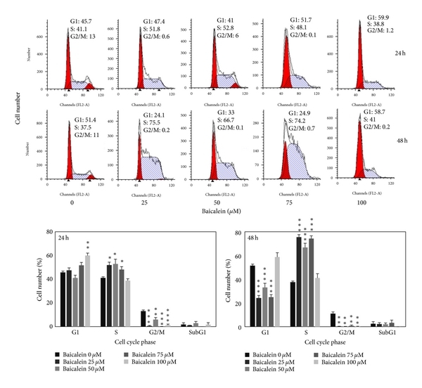
The cell cycle distribution changed by baicalein was analyzed by flow cytometric assay. Baicalein arrests cells in S phase after 24 h treatment at the concentration under 75 μM and in G1 phase at 100 μM. After treatment for 48 h, 100 μM baicalein continued to arrest cells in G1 without sub-G1 formation (Figure 2(a)). It suggests that baicalein, less than 50 μM, caused S phase arrest without significant cytotoxicity. One hundred μM baicalein arrested cells in G1 phase and induced cytotoxicity. One of G1/S transition promotion factors, cyclin D1, was dose-dependently decreased by baicalein (Figure 2(b)). It may contribute the reason to 100 μM baicalein-induced G1 arrest. Baicalein also decreased cyclin B1 expression dose-dependently (Figure 2(b)). Because cyclin B1 is an essential factor for entering G2/M phase, baicalein-decreased cyclin B1 may lead to S phase arrest. Baicalein immediately decreases cyclin D1 expression after treatment for 2 h (Figure 2(b)). It suggests that 100 μM baicalein effectively and quickly inhibits cell cycle progression at G1 phase.
579751.fig.002afig2
Figure 2: Effect of baicalein on cell cycle phase distribution in 5637 cells. (a) Baicalein induces cell cycle arrest. Cells were treated with vehicle or baicalein for 24 and 48 h, then were collected for cell cycle analysis (, , compared with vehicle). (b) Effect of baicalein on cyclin B1/D1 expression. Left, cells were treated with baicalein for 24 h. Right, cells were treated with 100 μM baicalein for 2, 6, 12, and 24 h.
3.3. Effect of Baicalein on the Regulation of Upstream Signal Factors

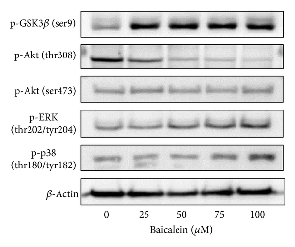
The intracellular signal factors p-GSK3β(ser9), p-AKT(thr308), p-AKT(ser473), p-ERK, and p-p38 were analyzed after baicalein treatment. The result of Figure 3(a) indicates that baicalein increases the phosphorylation of GSK3β(ser9), ERK(thr202/tyr204), and p38(thr180/tyr182). Though baicalein decreased the phosphorylation of AKT(thr308) and did not change p-AKT(ser473), the downstream GSK3β(ser9) was still phosphorylated at 24 h treatment. The time course of these baicalein-induced changes was also analyzed. As shown in Figure 3(b), baicalein inhibited p-AKT(thr308) phosphorylation from 2 h to 24 h; it suggests that baicalein inhibits Akt activity. GSK3β(ser9) was phosphorylated by baicalein from 2 h to 24 h, suggesting that baicalein also inhibits GSK3β activity. Both ERK and p38 pathways were early activated from 2 h to 24 h after baicalein treatment, ERK especially. The effect of baicalein on p65NF-κB was also analyzed. Without extracellular stimulation, most of p65NF-κB was found in cytoplasmic fraction in 5637 cells (Figure 3(c)). Baicalein dose-dependently inhibited the nuclear protein expression of p65NF-κB (Figure 3(c)). In summary, baicalein inhibits AKT and GSK3β activities, activates ERK and p38 pathways, and inhibits p65NF-κB-driven signals.
579751.fig.003afig3
Figure 3: Influence of baicalein on the signal pathways. (a) Effect of baicalein on the phosphorylation of GSK3β, AKT, ERK, and p38. 5637 cells were treated with 0.1% DMSO or baicalein for 24 h. (b) Time-course of baicalein-changed signal protein phosphorylation. 5637 cells treated with 100 μM baicalein or 0.1% DMSO for 2, 6, 12, and 24 h. The total cell lysates were extracted for western blot analysis. β-Actin was used as a loading control. (c) Effect of baicalein on the nuclear p65NF-κB expression. 5637 cells were treated with 0.1% DMSO or baicalein for 24 h. The cytoplasmic and nuclear extracts were prepared for Western blot analysis. α-Tubulin and Bub3 are the loading control of cytoplasmic and nuclear fraction, respectively.
3.4. Effect of Various Signal Protein Inhibitors on the Baicalein-Changed Cyclin B1/D1 Expression and Cell Viability

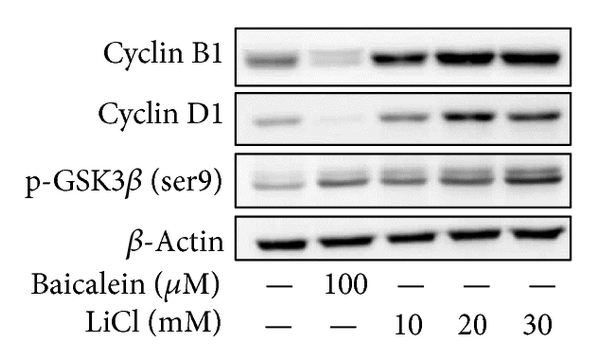
In order to understand the correlation between upstream signals and cyclin B1/D1 reduction, some specific inhibitors were used. Lithium chloride (LiCl) induces GSK3β(ser9) phosphorylation and inhibits GSK3βactivity [19, 20]. Baicalein or LiCl increased p-GSK3β(ser9), but only baicalein decreased cyclin B1/D1 expression (Figure 4(a)). It suggests that baicalein-inhibited cyclin B1/D1 expression is not mediated by GSK3βinhibition. On the contrary, LiCl dose-dependently increased cyclin B1/D1 expression, it suggests that baicalein-inhibited GSK3β pathway causes cyclin B1/D1 increase instead. LY294002, the inhibitor of PI3K-Akt pathway, inhibited the phosphorylation of AKT(ser473) but increased the phosphorylation of GSK3β(ser9) (Figure 4(b)). However, unlike baicalein, LY294002 did not reduce cyclin B1/D1 expression (Figure 4(b)). U0126, the inhibitors of MEK-ERK, slightly reversed baicalein-decreased cyclin B1 but not cyclin D1 (Figure 4(c)). The p38 kinase inhibitor SB203580 did not reverse baicalein-decreased cyclin B1/D1 expression (Figure 4(d)). Ro106-9920, an inhibitor of p65NF-κB, did not decrease cyclin B1/D1 expression (Figure 4(e)). These data indicate that baicalein-inhibited cyclin B1 is slightly mediated by ERK activation. The relationship of cell viability and baicalein-induced change in p-GSK3β(ser9), p-ERK, p-p38, and p65NF-κB was also analyzed. Using MTT assay (Figure 4(f)), MEK-ERK inhibitor U0126 and p38 kinase inhibitor SB203580 did not affect baicalein-reduced cell viability; the PI3K inhibitor LY294002 deteriorated baicalein-reduced cell viability; the p65NF-κB inhibitor Ro106-9920 reduced cell viability directly. It suggests that baicalein-induced p38, ERK, and GSK3β(ser9) phosphorylation does not play essential roles in cell growth inhibition. Only the baicalein-inhibited p65NF-κB activity leads to reduction of cell viability. In order to find out baicalein-reduced cyclin B1/D1 caused by de novo protein synthesis inhibition or proteasomal degradation stimulation, the translation inhibitor cycloheximide and the proteasome inhibitor MG132 were used for this study. After cyclohexamide treatment, baicalein did not reduce cyclin B1 anymore (Figure 4(g)). But baicalein still reduced cyclin D1 expression in the presence of cycloheximide or MG132 (Figure 4(g)). It suggests both de novo protein synthesis inhibition and proteasomal degradation stimulation are involved in baicalein-reduced cyclin D1 expression, and cyclin B1 decrease is only caused by de novo protein synthesis inhibition.
579751.fig.004afig4
Figure 4: Effect of various inhibitors on baicalein-reduced cyclin B1/D1 expression. (a–e) Effect of LiCl (a), LY294002 (b), U0126 (c), SB203580 (d), and Ro106-9920 (e) on baicalein-reduced cyclin B1/D1 expression. (f) Effect of various inhibitors on baicalein-inhibited cell viability. All above inhibitors were pretreated for 1 h and baicalein treatment for 24~72 h. The concentration of each chemicals: baicalein is 100 μM and others are 10 μM. (g) Effect of cycloheximide or MG132 on baicalein-reduced cyclin B1/D1 expression. Cycloheximide was pretreated for 30 min and baicalein treatment for 1 h in cyclin D1 detection and baicalein treatment for 6 h in cyclin B1 detection. MG132 was pretreated for 1 h and baicalein treatment for 6 h. The extracted cell lysates or nuclear proteins were analyzed by western blot.
3.5. Baicalein Blocks Migration and Invasion of 5637 Bladder Cancer Cells

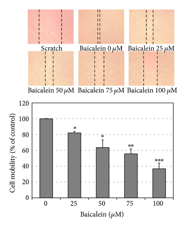
Using scratch assay, baicalein dose-dependently inhibited cell migration (Figure 5(a)). At 100 μM, baicalein shows 60% inhibition in cell migration, which is more effective than the inhibition in cell viability (33% inhibition at 100 μM, Figure 1(a)). By matrigel-coated invasion assay, baicalein also shows a significant inhibition dose-dependently (Figure 5(b)). On the other hand, baicalein reduced MMP2 and MMP9 mRNA expression (Figure 5(c)) and enzymatic activity (Figure 5(d)) in 5637 cells. It suggests that the baicalein-inhibited MMP2/9 activity may contribute its anti-migration and anti-invasion activity.
579751.fig.005afig5
Figure 5: Anti-migration and anti-invasion activities of baicalein in 5637 cells. (a) Baicalein inhibits 5637 cell migration. Bottom chart is the percentage of migrated cells as control is 100%. (b) Baicalein inhibits 5637 cell invasion. Bottom chart is the percentage of invaded cells as control is 100%. (c) Effect of baicalein on the mRNA expression of MMP-2 and MMP-9. Bottom charts are the quantitative results from three independent experiments. (d) Effect of baicalein on the activities of MMP-2 and MMP-9. The conditioned medium was collected 24 h after drug treatment. Twenty micrograms of protein obtained from the concentrated medium was analyzed by gelatin-zymography assay. (, , compared with vehicle).
3.6. Baicalein Slightly Inhibits Tumor Growth with Some Hepatotoxicity in a Mouse Orthotopic Bladder Tumor Model In Vivo

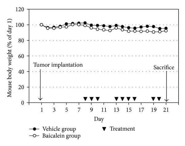
Based on the antigrowth and antimetastasis activity of baicalein in cell assay, the in vivo antitumor assay was analyzed. After bladder cell implantation on day 1, baicalein treatment started on day 8. The treatment did not show toxicity in appearance and body weight (Figure 6(a)). Baicalein did not significantly reduce bladder size, but the mean bladder volume was still reduced in baicalein-treated mice (from 49.5 mm3 to 35.9 mm3 in Figure 6(b)). The blood biochemical analysis shows no significant change in serum BUN and creatinine between control and baicalein treatment groups, a little increase in GPT value but without statistical significance, and a significant increase in serum GOT (Table 1). It suggests that baicalein treatment causes some hepatic toxicity in mice.

table1tab1
Table 1: Effect of vehicle and baicalein on the plasma biochemical parameters of mice at termination of treatment. Values are mean ± SE.
579751.fig.006afig6
Figure 6: The antitumor effect of baicalein in vivo. (a) Mouse body weight and drug schedule in the mouse orthotopic bladder tumor model. After MB49 cell implantation, baicalein or vehicle was applied by intraperitoneal injection. Mouse body weight was recorded every day. At the 21th day, the mice were sacrificed. (b) Effect of baicalein in reducing bladder tumor size. The bladder volume of each mouse was measured. There are 9 mice survived at the 21th day in vehicle group and 7 in baicalein group.
4. Discussion

This study provides some new information about baicalein used in the anticancer therapy. In cell study, baicalein decreases cyclin B1 protein expression through inhibiting de novo protein synthesis and inhibits cyclin D1 by inhibiting protein synthesis and promoting proteasomal degradation. Baicalein-inhibited cyclin B1 is partially mediated by ERK activation. Among the signal transduction molecules AKT, GSK3β, ERK, p38, and p65NF-κB, p65NF-κB inhibition plays the most important role in baicalein-reduced cell viability. In mouse orthotopic bladder tumor model, baicalein has a little inhibition effect on orthotopic bladder tumor growth but with some hepatic toxicity.

Baicalein produces different cytotoxicity in different cell lines. For example, it causes cell cycle arrest at G1 phase in breast cancer [21] and oral squamous cell carcinoma [22], at S phase in lung nonsmall carcinoma cell [23] and at G2/M phase in BFTC905 bladder cancer cells [9]. The differences may be caused by different doses and different cells used. In 5637 bladder cancer cells, baicalein arrests cells at S phase under 75 μM and at G1 phase at 100 μM without apoptotic cells (Figure 2(a)). In pancreatic carcinoma PaCa cells [11], bladder cancer BFTC905 cells [9], and colorectal carcinoma HCT116 cells [10], baicalein induces cell apoptotic death at the dose between 5 to 60 μM. Baicalein also has a wide range on cytotoxicity of different cell lines, the IC50 is under 20 μM in gastric cancer cells AGS and MKN-28 [24], prostate carcinoma LNCaP [25], and JCA-1 [26], between 20 to 50 μM in leukemia HL-60 [27], bladder cancer BFTC905 [9], hepatic cancer Hep G2 [28], and myeloma cell U266 [29], and more than 100 μM in 5637 bladder cancer (Figure 1(a)), oral squamous carcinoma HSC-3 [22], leukemia THP-1, and osteogenic cancer cell HOS [30]. Although the detail mechanisms about the wide-range cytotoxicity are still unclear, this property may provide a specific and lower hazard anticancer effect for the higher sensitive tumors.

PI3K, the upstream signal of AKT, has been reported to be inhibited by baicalein [31]. In our study, baicalein inhibits pAKT(thr308) phosphorylation and has no influence on pAKT(ser473) (Figure 2(b)). The pAKT inhibition phenomenon also has been reported in prostate cancer cell DU145 [32] and oral squamous carcinoma HSC-3 [22]. On the other hand, the pAKT(ser473) activation has been reported in bladder cancer BFTC905 [9, 10]. Because the pAKT(thr308) phosphorylation site is the direct target site for PI3K-PDK1 [33], it will be downregulated after PI3K inhibition by baicalein (Figure 4(b)). The ser473 site of AKT is phosphorylated by rictor-mTOR [34], not PI3K; therefore, it may be the reason for the no effect of baicalein on the phosphorylation of pAKT(ser473). Even though the pAKT(thr308) is decreased by baicalein, pGSK3β(ser9), one AKT downstream [35], is still phosphorylated by baicalein (Figure 3(b)). Because the phosphorylation of pGSK3β(ser9) is achieved by numerous kinases, not only AKT [35], baicalein-induced pGSK3β(ser9) may be caused by the influence of other kinase(s).

NF-κB, an important inflammatory transcription factor, is inhibited by baicalein in 5637 cells (Figure 3(c)). Baicalein-inhibited p65NF-κB activation has also been reported in human mast cells [36], mouse macrophages [37, 38], human myeloma cells [29], and brain microglia [39, 40]. According to the important role of p65NF-κB in tumor progression and metastasis [41, 42], the inhibition of baicalein on nuclear NF-κB is a critical function in its anti-inflammation and anticancer application. In human hepatoma cells, baicalein shows anti-migration property with NF-κB inhibition [12]. There is one report indicates that GSK3β inhibition results in inhibiting NF-κB activity [43]; therefore, the mechanism of baicalein-inhibited p65NF-κB activity may be partially mediated by baicalein-inhibited GSK3β. In Figure 4(f), among the 4 signal inhibitors, the cell viability decreases at most by NF-κB inhibitor Ro106-9920, which indicates that NF-κB is a critical factor for proliferation of 5637 cells.

Baicalein inhibits the protein expression of cyclin B1 [9, 23, 44] and cyclin D1 [22, 32, 44], has also been reported by some studies, but the mechanism is still unclear. In this study, we first suggest that baicalein decreases cyclin B1 expression through inhibiting de novo protein synthesis but not promoting proteasomal degradation and decreases cyclin D1 by both ways (Figure 4(g)). On the other hand, cyclin B1 reduction is partially mediated by ERK activation (Figure 4(c)). Luteolin, a natural flavonoid with structure similar to baicalein, decreases cyclin D1 expression by increasing proteasomal degradation [45]. Though the structures are similar between baicalein and luteolin, the mechanisms for cyclin D1 reduction are different. Luteolin enhances proteasomal degradation via decreasing GSK3β(ser9) phosphorylation, but baicalein increases GSK3β(ser9) phosphorylation (Figures 3(a) and 3(b)). Therefore, there is (are) other pathway(s) for inducing cyclin D1 degradation by baicalein.

In addition to anti-proliferation, baicalein also inhibits cancer cell metastasis. Either in scratch assay (Figure 5(a)) or in Matrigel-coated transwell assay (Figure 5(b)), they point out the anti-migration and anti-invasion property of baicalein. In this study, we confirm this inhibition, like others [12, 13], mediated by inhibiting MMP-2/9 activities (Figure 5(d)). The correlated signal pathways need to be further investigated. In the orthotopic bladder tumor model, baicalein shows a little effect on inhibiting bladder tumor growth (Figure 6). One report indicates that baicalein significantly reduces tumor volume in a nude mice model [12]. Comparing these two animal models, we use higher dose and lower frequency of baicalein, which may result in the lower efficiency. But it still notices that baicalein induces hepatic toxicity with GOT value increase. In order to avoid hepatic toxicity, it is better to use baicalein locally, for example, by intravesical application for bladder tumor therapy.

5. Conclusions

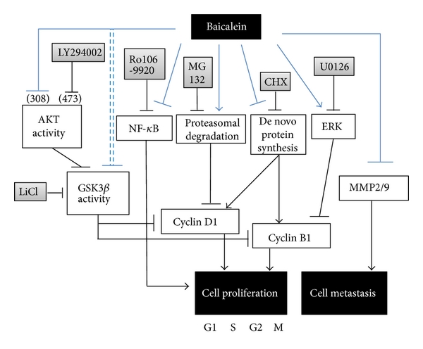
In this study, baicalein decreases cyclin D1 protein expression through inhibiting de novo protein synthesis and promoting proteasomal degradation and decreases cyclin B1 by inhibiting de novo protein synthesis. Baicalein-inhibited cyclin B1 expression is slightly mediated by ERK activation. The mechanism of baicalein in anti-proliferation and anti-metastasis is concluded in Figure 7. Among the signal transduction molecules AKT, GSK3β, ERK, p38, and p65NF-κB, p65NF-κB inhibition plays the most important role in baicalein-reduced cell viability. In mouse orthotopic bladder tumor model, baicalein has a little effect on orthotopic bladder tumor growth inhibition but with some hepatic toxicity.
579751.fig.007fig7
Figure 7: The pathway scheme of baicalein on the cell proliferation inhibition and cell metastasis inhibition in human bladder cancer cell 5637. The anti-proliferation and anti-metastasis activities of baicalein may contribute the anti-bladder-tumor effect in vivo.
Abbreviations

FBS: Fetal bovine serum
MMP-2: Matrix metalloproteinase-2
MMP-9: Matrix metalloproteinase-9
MTT: 3-(4,5-dimethylthiazol-2-yl)2,5-diphenyltetrazolium bromide
NF-κB: Nuclear factor-kappaB
PI3K: Phosphatidylinositol 3-kinase.
Conflict of Interests

The authors declare that there is no conflict of interests.

Authors’ Contribution

J.-Y. Wu and K.-W. Tsai contributed equally to this work.

Acknowledgments

This work is supported by Grants from the National Science Council NSC101-2320-B-415-002-MY3 of the Republic of China and Buddhist Tzuchi Dalin General Hospital, Dalin Town, Chiayi, Taiwan.

References

1 A. Jemal, F. Bray, M. M. Center, J. Ferlay, E. Ward, and D. Forman, “Global cancer statistics,” CA Cancer Journal for Clinicians, vol. 61, no. 2, pp. 69–90, 2011. View at Publisher · View at Google Scholar · View at Scopus
2 R. Siegel, D. Naishadham, and A. Jemal, “Cancer statistics 2012,” A Cancer Journal for Clinicians, vol. 62, no. 1, pp. 10–29, 2012.
3 D. J. McConkey, S. Lee, W. Choi et al., “Molecular genetics of bladder cancer: emerging mechanisms of tumor initiation and progression,” Urologic Oncology, vol. 28, no. 4, pp. 429–440, 2010. View at Publisher· View at Google Scholar · View at Scopus
4 X. R. Wu, “Urothelial tumorigenesis: a tale of divergent pathways,” Nature Reviews Cancer, vol. 5, no. 9, pp. 713–725, 2005. View at Publisher · View at Google Scholar · View at Scopus
5 L. C. Pagliaro and P. Sharma, “Review of metastatic bladder cancer,” Minerva Urologica e Nefrologica, vol. 58, no. 1, pp. 53–71, 2006. View at Scopus
6 C. C. Lin and D. E. Shieh, “The anti-inflammatory activity of Scutellaria rivularis extracts and its active components, baicalin, baicalein and wogonin,” American Journal of Chinese Medicine, vol. 24, no. 1, pp. 31–36, 1996. View at Scopus
7 Z. H. Shao, T. L. Vanden Hoek, Y. Qin et al., “Baicalein attenuates oxidant stress in cardiomyocytes,” American Journal of Physiology, vol. 282, no. 3, pp. H999–H1006, 2002. View at Scopus
8 L. G. Chen, L. Y. Hung, K. W. Tsai et al., “Wogonin, a bioactive flavonoid in herbal tea, inhibits inflammatory cyclooxygenase-2 gene expression in human lung epithelial cancer cells,” Molecular Nutrition and Food Research, vol. 52, no. 11, pp. 1349–1357, 2008. View at Publisher · View at Google Scholar · View at Scopus
9 J. I. Chao, W. C. Su, and H. F. Liu, “Baicalein induces cancer cell death and proliferation retardation by the inhibition of CDC2 kinase and survivin associated with opposite role of p38 mitogen-activated protein kinase and AKT,” Molecular Cancer Therapeutics, vol. 6, no. 11, pp. 3039–3048, 2007. View at Publisher · View at Google Scholar · View at Scopus
10 R. H. Jiang, W. C. Su, H. F. Liu, H. S. Huang, and J. I. Chao, “Opposite expression of securin and γ-H2AX regulates baicalein-induced cancer cell death,” Journal of Cellular Biochemistry, vol. 111, no. 2, pp. 274–283, 2010. View at Publisher · View at Google Scholar · View at Scopus
11 H. Takahashi, M. C. Chen, H. Pham et al., “Baicalein, a component of Scutellaria baicalensis, induces apoptosis by Mcl-1 down-regulation in human pancreatic cancer cells,” Biochimica et Biophysica Acta, vol. 1813, no. 8, pp. 1465–1474, 2011. View at Publisher · View at Google Scholar · View at Scopus
12 Y. W. Chiu, T. H. Lin, W. S. Huang, et al., “Baicalein inhibits the migration and invasive properties of human hepatoma cells,” Toxicology and Applied Pharmacology, vol. 255, no. 3, pp. 316–326, 2011.
13 L. Wang, Y. Ling, Y. Chen et al., “Flavonoid baicalein suppresses adhesion, migration and invasion of MDA-MB-231 human breast cancer cells,” Cancer Letters, vol. 297, no. 1, pp. 42–48, 2010. View at Publisher · View at Google Scholar · View at Scopus
14 B. Wu, J. Li, D. Huang, et al., “Baicalein mediates inhibition of migration and invasiveness of skin carcinoma through Ezrin in A431 cells,” BMC Cancer, vol. 11, article 527, 2011.
15 J. T. Fox, S. Sakamuru, R. Huang, et al., “High-throughput genotoxicity assay identifies antioxidants as inducers of DNA damage response and cell death,” Proceedings of the National Academy of Sciences of the United States of America, vol. 109, no. 14, pp. 5423–5428, 2012.
16 J. Y. Wu, K. T. Chung, Y. W. Liu et al., “Synthesis and biological evaluation of novel C(6) modified baicalein derivatives as antioxidative agents,” Journal of Agricultural and Food Chemistry, vol. 56, no. 8, pp. 2838–2845, 2008. View at Publisher · View at Google Scholar · View at Scopus
17 C. H. Shen, J. J. Shee, J. Y. Wu, Y. W. Lin, J. D. Wu, and Y. W. Liu, “Combretastatin A-4 inhibits cell growth and metastasis in bladder cancer cells and retards tumour growth in a murine orthotopic bladder tumour model,” British Journal of Pharmacology, vol. 160, no. 8, pp. 2008–2027, 2010. View at Publisher · View at Google Scholar · View at Scopus
18 S. Y. Wu, Y. R. Lee, C. C. Huang, et al., “Curcumin-induced heme oxygenase-1 expression plays a negative role for its anti-cancer effect in bladder cancers,” Food and Chemical Toxicology, vol. 50, no. 10, pp. 3530–3536, 2012.
19 P. S. Klein and D. A. Melton, “A molecular mechanism for the effect of lithium on development,” Proceedings of the National Academy of Sciences of the United States of America, vol. 93, no. 16, pp. 8455–8459, 1996. View at Publisher · View at Google Scholar · View at Scopus
20 F. Zhan, C. J. Phiel, L. Spece, N. Gurvich, and P. S. Klein, “Inhibitory phosphorylation of glycogen synthase kinase-3 (GSK-3) in response to lithium: evidence for autoregulation of GSK-3,” Journal of Biological Chemistry, vol. 278, no. 35, pp. 33067–33077, 2003. View at Publisher · View at Google Scholar· View at Scopus
21 Q. M. Zhou, S. Wang, H. Zhang et al., “The combination of baicalin and baicalein enhances apoptosis via the ERK/p38 MAPK pathway in human breast cancer cells,” Acta Pharmacologica Sinica, vol. 30, no. 12, pp. 1648–1658, 2009. View at Publisher · View at Google Scholar · View at Scopus
22 Y. H. Cheng, L. A. Li, P. Lin, et al., “Baicalein induces G1 arrest in oral cancer cells by enhancing the degradation of cyclin D1 and activating AhR to decrease Rb phosphorylation,” Toxicology and Applied Pharmacology, vol. 263, no. 3, pp. 360–367, 2012.
23 H. W. C. Leung, W. H. Yang, M. Y. Lai, C. J. Lin, and H. Z. Lee, “Inhibition of 12-lipoxygenase during baicalein-induced human lung nonsmall carcinoma H460 cell apoptosis,” Food and Chemical Toxicology, vol. 45, no. 3, pp. 403–411, 2007. View at Publisher · View at Google Scholar · View at Scopus
24 B. C. Y. Wong, W. P. Wang, C. H. Cho et al., “12-Lipoxygenase inhibition induced apoptosis in human gastric cancer cells,” Carcinogenesis, vol. 22, no. 9, pp. 1349–1354, 2001. View at Scopus
25 M. Bonham, J. Posakony, I. Coleman, B. Montgomery, J. Simon, and P. S. Nelson, “Characterization of chemical constituents in Scutellaria baicalensis with antiandrogenic and growth-inhibitory activities toward prostate carcinoma,” Clinical Cancer Research, vol. 11, no. 10, pp. 3905–3914, 2005. View at Publisher · View at Google Scholar · View at Scopus
26 S. Chen, Q. Ruan, E. Bedner et al., “Effects of the flavonoid baicalin and its metabolite baicalein on androgen receptor expression, cell cycle progression and apoptosis of prostate cancer cell lines,” Cell Proliferation, vol. 34, no. 5, pp. 293–304, 2001. View at Publisher · View at Google Scholar · View at Scopus
27 M. K. Roy, K. Nakahara, V. Na Thalang et al., “Baicalein, a flavonoid extracted from a methanolic extract of Oroxylum indicum inhibits proliferation of a cancer cell line in vitro via induction of apoptosis,” Pharmazie, vol. 62, no. 2, pp. 149–153, 2007. View at Publisher · View at Google Scholar · View at Scopus
28 W. H. Chang, C. H. Chen, and F. J. Lu, “Different effects of baicalein, baicalin and wogonin on mitochondrial function, glutathione content and cell cycle progression in human hepatoma cell lines,” Planta Medica, vol. 68, no. 2, pp. 128–132, 2002. View at Publisher · View at Google Scholar · View at Scopus
29 Z. Ma, K. I. Otsuyama, S. Liu et al., “Baicalein, a component of Scutellaria radix from Huang-Lian-Jie-Du-Tang (HLJDT), leads to suppression of proliferation and induction of apoptosis in human myeloma cells,” Blood, vol. 105, no. 8, pp. 3312–3318, 2005. View at Publisher · View at Google Scholar · View at Scopus
30 M. Himeji, T. Ohtsuki, H. Fukazawa et al., “Difference of growth-inhibitory effect of Scutellaria baicalensis-producing flavonoid wogonin among human cancer cells and normal diploid cell,” Cancer Letters, vol. 245, no. 1-2, pp. 269–274, 2007. View at Publisher · View at Google Scholar · View at Scopus
31 D. Kong, Y. Zhang, T. Yamori, H. Duan, and M. Jin, “Inhibitory activity of flavonoids against class I phosphatidylinositol 3-kinase isoforms,” Molecules, vol. 16, no. 6, pp. 5159–5167, 2011. View at Publisher· View at Google Scholar · View at Scopus
32 G. P. Pidgeon, M. Kandouz, A. Meram, and K. V. Honn, “Mechanisms controlling cell cycle arrest and induction of apoptosis after 12-lipoxygenase inhibition in prostate cancer cells,” Cancer Research, vol. 62, no. 9, pp. 2721–2727, 2002. View at Scopus
33 L. Stephens, K. Anderson, D. Stokoe et al., “Prohtein kinase B kinases that mediate phosphatidylinositol 3,4,5-trisphosphate-dependent activation of protein kinase B,” Science, vol. 279, no. 5351, pp. 710–714, 1998. View at Publisher · View at Google Scholar · View at Scopus
34 D. D. Sarbassov, D. A. Guertin, S. M. Ali, and D. M. Sabatini, “Phosphorylation and regulation of Akt/PKB by the rictor-mTOR complex,” Science, vol. 307, no. 5712, pp. 1098–1101, 2005. View at Publisher · View at Google Scholar · View at Scopus
35 R. S. Jope, C. J. Yuskaitis, and E. Beurel, “Glycogen synthase kinase-3 (GSK3): inflammation, diseases, and therapeutics,” Neurochemical Research, vol. 32, no. 4-5, pp. 577–595, 2007. View at Publisher · View at Google Scholar · View at Scopus
36 C. J. Hsieh, K. Hall, T. Ha, C. Li, G. Krishnaswamy, and D. S. Chi, “Baicalein inhibits IL-1β- and TNF-α-induced inflammatory cytokine production from human mast cells via regulation of the NF-κB pathway,” Clinical and Molecular Allergy, vol. 5, article 5, 2007. View at Publisher · View at Google Scholar · View at Scopus
37 B. Y. Kang, S. W. Chung, S. H. Kim, D. Cho, and T. S. Kim, “Involvement of nuclear factor-κB in the inhibition of interleukin-12 production from mouse macrophages by baicalein, a flavonoid in Scutellaria baicalensis,” Planta Medica, vol. 69, no. 8, pp. 687–691, 2003. View at Publisher · View at Google Scholar · View at Scopus
38 M. H. Kim, S. Y. Ryu, M. A. Bae, J. S. Choi, Y. K. Min, and S. H. Kim, “Baicalein inhibits osteoclast differentiation and induces mature osteoclast apoptosis,” Food and Chemical Toxicology, vol. 46, no. 11, pp. 3375–3382, 2008. View at Publisher · View at Google Scholar · View at Scopus
39 C. J. Chen, S. L. Raung, S. L. Liao, and S. Y. Chen, “Inhibition of inducible nitric oxide synthase expression by baicalein in endotoxin/cytokine-stimulated microglia,” Biochemical Pharmacology, vol. 67, no. 5, pp. 957–965, 2004. View at Publisher · View at Google Scholar · View at Scopus
40 K. Suk, H. Lee, S. S. Kang, G. J. Cho, and W. S. Choi, “Flavonoid baicalein attenuates activation-induced cell death of brain microglia,” Journal of Pharmacology and Experimental Therapeutics, vol. 305, no. 2, pp. 638–645, 2003. View at Publisher · View at Google Scholar · View at Scopus
41 J. I. Inoue, J. Gohda, T. Akiyama, and K. Semba, “NF-κB activation in development and progression of cancer,” Cancer Science, vol. 98, no. 3, pp. 268–274, 2007. View at Publisher · View at Google Scholar · View at Scopus
42 G. Sethi, B. Sung, and B. B. Aggarwal, “Nuclear factor-κB activation: from bench to bedside,” Experimental Biology and Medicine, vol. 233, no. 1, pp. 21–31, 2008. View at Publisher · View at Google Scholar · View at Scopus
43 J. Deng, W. Xia, S. A. Miller, Y. Wen, H. Y. Wang, and M. C. Hung, “Crossregulation of NF-κB by the APC/GSK-3β/β-catenin pathway,” Molecular Carcinogenesis, vol. 39, no. 3, pp. 139–146, 2004. View at Publisher · View at Google Scholar · View at Scopus
44 H. Z. Lee, H. W. C. Leung, M. Y. Lai, and C. H. Wu, “Baicalein induced cell cycle arrest and apoptosis in human lung squamous carcinoma CH27 cells,” Anticancer Research, vol. 25, no. 2, pp. 959–964, 2005.View at Scopus
45 C. S. Ong, J. Zhou, C. N. Ong, and H. M. Shen, “Luteolin induces G1 arrest in human nasopharyngeal carcinoma cells via the Akt-GSK-3β-cyclin D1 pathway,” Cancer Letters, vol. 298, no. 2, pp. 167–175, 2010. View at Publisher · View at Google Scholar · View at Scopus

δ-Elemene (delta Elemene)

Cancer: Lung
Action: Induces apoptosis, inhibits NF-kappa B
δ-elemene significantly induced apoptosis of NCI-H292, as shown by 3-(4,5-dimethylthiazol-2-yl)-2,5-diphenyltetrazolium bromide (MTT) assay, DNA fragmentation measurement, Annexin V (AnV) binding of externalized phosphatidylserine and the mitochondrial probe JC-1 using flow cytometry.

Treatment of NCI-H292 with δ-elemene increased both p38 mitogen-activated protein kinase (MAPK) and inducible nitric oxide synthese (iNOS) levels, suggesting these two molecules maybe relate to the apoptotic effect of δ-elemene. The cells with Bcl-2 or Bcl-xL over-expression showed an elevation of nuclear factor kappa B (NF-kappa B) activity, accompanying a significant reduction of δ-elemene-induced apoptosis.

Furthermore, inhibition of NF-kappa B by IkBαSR, which is a powerful inhibitor of NF-kappa B, restored the ability of δ-elemene to induce apoptosis in the cells transfected with Bcl-2. These data strongly indicated that the apoptotic effect of δ-elemene on NCI-H292 was closely associated with the activity of NF-kappa B, which was up-regulated by Bcl-2 and Bcl-xL.
In conclusion, δ-elemene induced apoptosis in NCI-H292 cells. The apoptotic effect of δ-elemene could be significantly offset by over-expression of either Bcl-2 or Bcl-xL. Bcl-2 and Bcl-xL were able to increase the activity of NF-kappa B, which was a known anti-apoptotic molecule in human lung cancer cells.

Source
Xie CY, Yang W, Ying J, et al. B-cell lymphoma-2 over-expression protects δ-elemene-induced apoptosis in human lung carcinoma mucoepidermoid cells via a nuclear factor kappa B-related pathway. Biol Pharm Bull. 2011;34(8):1279-86.

Coriolus versicolor (yun zhi)

Cancer: Breast, gastric, colorectal
Action: Chemotherapy adjunct, survival

Systematic review and meta-analysis technique were used to aggregate and analyze the efficacy of Yun Zhi on survival in cancer patients from 13 clinical trials using computerized database and manual search. The findings show that Yun Zhi results in a significant survival advantage compared with standard conventional anti-cancer treatment alone. Of patient randomized to Yun Zhi, there was a 9% absolute reduction in 5-year mortality, resulting in one additional patient alive for every 11 patients treated. In patients with breast cancer, gastric cancer, or colorectal cancer treated with chemotherapy, the effects of the combination of Yun Zhi preparation on the overall 5-year survival rate was more evident, but not in esophageal cancer and nasophayngeal carcinoma. However, subgroup analysis could not conclude which type of anti-cancer treatment may maximize the benefit from Yun Zhi.

This meta-analysis has provided strong evidence that Yun Zhi would have survival benefit in cancer patients, particularly in carcinoma of breast, gastric and colorectal. Nevertheless, the findings highlight the need for further evidence from prospective studies of outcome to guide future potential modifications of treatment regimes.

Source
Eliza WL, Fai CK, Chung LP. Efficacy of Yun Zhi (Coriolus versicolor) on survival in cancer patients: systematic review and meta-analysis. Recent Pat Inflamm Allergy Drug Discov. 2012 Jan;6(1):78-87.

Oridonin phosphate

Cancer: Breast

Action: Induces apoptosis, up-regulates Bax and down-reguates Bcl-2, induces autophagy

Oridonin phosphate is one of the derivatives of oridonin. The results by Li et al., (2015) demonstrated that oridonin phosphate inhibited the proliferation of MDA-MB-436 and MDA-MB-231 cells in a dose- and time-dependent manner. Cell apoptosis rate was detected in oridonin phosphate-treated breast cancer cells and the data demonstrated that oridonin phosphate induced cell apoptosis of breast cancer cells in time- and dose-dependent manner.

The results showed that the expression level of Bax was up-regulated and the expression level of Bcl-2 was down-regulated. Meanwhile, the level of cleaved caspase-9 was significantly increased when the cells were treated with 40 μM of oridonin phosphate for 48 h, although the expression level of pro-caspase-9 was not obviously changed. All of the data revealed that mitochondrial apoptosis pathway may be involved in the cell apoptosis induced by oridonin phosphate in breast cancer cells.

Additionally, they further explored the relationship between apoptosis and autophagy specifically induced by oridonin phosphate in breast cancer cells. Taken together, the compound of oridonin phosphate simultaneously induced cell apoptosis and autophagy in breast cancer cells.

Inhibition oridonin phosphate-induced cell autophagy suppressed the progression of cell apoptosis, which revealed that oridonin phosphate-induced autophagy participated in up-regulation of apoptosis in human breast cancer cells.

Source
Li Y, Wang Y, Wang S, Gao Y, Zhang X, Lu C. Oridonin phosphate-induced autophagy effectively enhances cell apoptosis of human breast cancer cells. Med Oncol. 2015 Jan;32(1):365. doi: 10.1007/s12032-014-0365-1.

Berberine

Cancer: Multiple Myeloma

Action: Down-regulates miR-21 levels through IL6/STAT3

Berberine is known to modulate microRNA (miRNA) levels, although the mechanism for this action is unknown. Luo et al. previously demonstrate that the expression of 87 miRNAs is differentially affected by berberine in multiple myeloma cells. Among 49 miRNAs that are down-regulated, nine act as oncomirs, including miR-21. Integrative analysis showed that 28 of the down-regulated miRNAs participate in tumor protein p53 (TP53) signaling and other cancer pathways. miR-21 is involved in all these pathways, and is one of the most important oncomirs to be affected by berberine in multiple myeloma cells.

They confirmed that berberine down-regulated miRNA-21 expression and significantly up-regulated the expression of programmed cell death 4 (PDCD4), a predicted miR-21 target. Depletion of PDCD4 by short interfering RNA could rescue berberine-induced cytotoxicity in multiple

Results suggest that berberine suppresses multiple myeloma cell growth, at least in part, by down-regulating miR-21 levels possibly through IL6/STAT3. This led to increased PDCD4 expression, which is likely to result in suppression of the p53 signaling pathway.

Source

Luo X, Gu J, Zhu R, et al. Integrative analysis of differential miRNA and functional study of miR-21 by seed-targeting inhibition in multiple myeloma cells in response to berberine. BMC Syst Biol. 2014 Jul 7;8:82. doi: 10.1186/1752-0509-8-82


Cancer: Multiple Myeloma

Action: Triggers hypomethylation

Berberine reduces the proliferation and induces apoptosis in the multiple myeloma cell line, U266.Qing et al., (2014) explored the detailed mechanism by analysing the gene expression profiles in U266 treated with or without berberine. DNMT1 andDNMT3B, encoding for a highly conserved member of the DNA methyltransferases, decreased significantly. Results show that berberine can repress the expression of DNMT1 and DNMT3B, which triggers hypomethylation of TP53 by changing the DNA methylation level and the alteration of p53 dependent signal pathway in human multiple melanoma cell U266.

Source

Qing Y, Hu H, Liu Y, Feng T, Meng W, Jiang L, Sun Y, Yao Y. Berberine induces apoptosis in human multiple myeloma cell line U266 through hypomethylation of p53 promoter. Cell Biol Int. 2014 May;38(5):563-70.

Emodin

Cancer: Multiple Myeloma

Action: Inhibits interleukin-6, activates of caspase-3 and caspase-9

Muto et al., (2007) show that emodin significantly induces cytotoxicity in the human myeloma cells through the elimination of myeloid cell leukemia 1 (Mcl-1). Emodin inhibited interleukin-6-induced activation of Janus-activated kinase 2 (JAK2) and phosphorylation of signal transducer and activator of transcription 3 (STAT3), followed by the decreased expression of Mcl-1. Activation of caspase-3 and caspase-9 was triggered by emodin, but the expression of other antiapoptotic Bcl-2 family members, except Mcl-1, did not change in the presence of emodin. To clarify the importance of Mcl-1 in emodin-induced apoptosis, the Mcl-1 expression vector was introduced into the human myeloma cells by electroporation. Induction of apoptosis by emodin was almost abrogated in Mcl-1-overexpressing myeloma cells as the same level as in parental cells, which were not treated with emodin. In conclusion, emodin inhibits interleukin-6-induced JAK2/STAT3 pathway selectively and induces apoptosis in myeloma cells via down-regulation of Mcl-1, which is a good target for treating myeloma. Taken together, our results show emodin as a new potent anticancer agent for the treatment of multiple myeloma patients.

Source

Muto A, Hori M, Sasaki Y, et al. Emodin has a cytotoxic activity against human multiple myeloma as a Janus-activated kinase 2 inhibitor. Mol Cancer Ther. 2007 Mar;6(3):987-94.

Cancer: Multiple myeloma

Action: Induces apoptosis

Although novel drugs have contributed immensely to improving outcomes of patients with multiple myeloma (MM), many patients develop drug resistance and ultimately succumb to MM. Papanikolaou et al., (2014) show that artesunate, an anti-malarial drug, reliably induces cell death in vitro in naïve as well as drug-resistant MM cells at concentrations shown to be safe in humans. Artesunate induced apoptosis predominantly through the non-caspase mediated pathway by primarily targeting mitochondria and causing outer mitochondrial membrane permeabilization that led to cytosolic and subsequent nuclear translocation of mitochondrial proteins apoptosis inducing factor (AIF) and endonuclease G (EndoG). These effects were present before apoptosis was evident and were related to intracellular levels of bivalent iron (Fe+2). Artesunate’s unique mechanism probably was at least partially responsible for, its ability to act synergistically with multiple anti-myeloma agents. Their findings suggest that artesunate acts through iron to affect the mitochondria and induce low ROS and non-caspase-mediated apoptosis. Its potency, toxicity profile, and synergism with other drugs make it an intriguing new candidate for MM treatment.

Source

Papanikolaou X, Johnson S, Garg T et al. Artesunate overcomes drug resistance in multiple myeloma by inducing mitochondrial stress and non-caspase apoptosis. Oncotarget. 2014 Jun 30;5(12):4118-28.

(DCIS) Ductal Cell Carcinoma in Situ

(DCIS) Ductal Cell Carcinoma in Situ

Action: inhibits and reverses precancerous lesions of breast cancer
inhibits ki67 and mRNA expression

Yanghe Huayan Decoction (YHD) Cornu Cervi Degelatinatium (lu jiao shuang) 12 g, prepared rhizome of rehmannia (shu di) 9 g, Cortex Cinnamomi (rou gui) 6 g, Semen Sinapis (bai jie zi) 3 g, Radix Curcumae (yu jin) 12 g, Psuedobulbus Cremastrae (shan ci gu) 15 g, Bulbus Frittillariae Thunbergii (zhe bei mu) 9 g, licorice root (gan cao) 6 g at the daily dose of 7.2 g/kg.

YHD could partially inhibit and reverse canceration of DCIS. It also could inhibit ki67 protein and mRNA expression. Its effect was similar to tamoxifen.

It is suitable for prevention and treatment of precancerous lesions of breast cancer (Li et al., 2014).

References


Li JW, Liu XF, Chen HZ, Chen HH, Shi GX, Wang SJ. [Effect of yanghe huayan decoction on precancerosis of breast cancer, protein and mRNA expression of ki67: an experimental research]. Zhongguo Zhong Xi Yi Jie He Za Zhi. 2014 Aug;34(8):970-5. [Article in Chinese]

Cancer: Tongue

Berberine

Cancer: Tongue

Action: Down-regulates u-PA, MMP-2 and -9 expressions

There is increasing evidence that urokinase-type plasminogen activator (u-PA) and matrix metalloproteinases (MMPs) play an important role in cancer metastasis and angiogenesis. Inhibition of u-PA and MMPs could suppress migration and invasion of cancer cells. Berberine, one of the main constituents of the plant Rhizoma coptidis, is a type of isoquinoline alkaloid, reported to have anti-cancer effects in different human cancer cell lines.

Here, we report that berberine inhibited migration and invasion of human SCC-4 tongue squamous carcinoma cells. This action was mediated by the p-JNK, p-ERK, p-p38, IkappaK and NF-kappaB signaling pathways resulting in inhibition of MMP-2 and -9 in human SCC-4 tongue squamous carcinoma cells. Analysis also showed that berberine inhibited the levels of urokinase-plasminogen activator (u-PA). These results suggest that berberine down-regulates u-PA, MMP-2 and -9 expressions in SCC-4 cells through the FAK, IKK and NF-kappaB mediated pathways and a novel function of berberine is to inhibit the invasive capacity of malignant cells.

Reference

Ho YT, Yang JS, Li TC, Lin JJ, et al. Berberine suppresses in vitro migration and invasion of human SCC-4 tongue squamous cancer cells through the inhibitions of FAK, IKK, NF-kappaB, u-PA and MMP-2 and -9. Cancer Lett. 2009 Jul 8;279(2):155-62. doi: 10.1016/j.canlet.2009.01.033.

Gypenosides

Cancer: Oral

Action: Downregulates NF-κB, COX-2, extracellular signal-regulated kinase 1/2 (ERK1/ 2), matrix metalloproteinase-9, -2 (MMP-9, -2), sevenless homolog (SOS), Ras, urokinase-type plasminogen activator (uPA), focal adhesion kinase (FAK) and RAC-alpha serine/threonine-protein kinase (Akt)

Gypenosides (Gyp), found in Gynostemma pentaphyllum Makino, has been used as a folk medicine in the Chinese population for centuries and is known to have diverse pharmacologic effects, including anti-proliferative and anti-cancer actions. However, the effects of Gyp on prevention from invasion and migration of oral cancer cells are still unsatisfactory.

The purpose of this study was to investigate effects of Gyp treatment on migration and invasion of SAS human oral cancer cells. SAS cells were cultured in the presence of 90 and 180 μg/mL Gyp for 24 and 48 hours. Gyp induced cytotoxic effects and inhibited SAS cells migration and invasion in dose- and time-dependent response.

Gyp decreased the abundance of several proteins, including nuclear factor-kappa B (NF-κB), cyclooxygenase-2 (COX-2), extracellular signal-regulated kinase 1/2 (ERK1/ 2), matrix metalloproteinase-9, -2 (MMP-9, -2), sevenless homolog (SOS), Ras, urokinase-type plasminogen activator (uPA), focal adhesion kinase (FAK) and RAC-alpha serine/threonine-protein kinase (Akt), in a time-dependent manner. The inhibition of NF-κB and MMP-2, -7 and -9 signaling may be one of the mechanisms that is present in Gyp-inhibited cancer cell invasion and migration.

Reference

Lu KW, Chen JC, Lai TY, Yang JS, et al. Gypenosides inhibits migration and invasion of human oral cancer SAS cells through the inhibition of matrix metalloproteinase-2 -9 and urokinase-plasminogen by ERK1/2 and NF-kappa B signaling pathways. Hum Exp Toxicol. 2011 May;30(5):406-15. doi: 10.1177/0960327110372405

Emodin

Cancer: Tongue

Action: Downregulates MMP-2, u-PA

Emodin, aloe-emodin and rhein are major compounds in rhubarb (Rheum palmatum L.), used in Chinese herbal medicine, and found to have antitumor properties including cell cycle arrest and apoptosis in many human cancer cells. Our previous studies also showed that emodin, aloe-emodin and rhein induced apoptosis in human tongue cancer SCC-4 cells. In the present study, we investigated whether or not emodin, aloe-emodin and rhein inhibited migration and invasion of SCC-4 cells.

Herein, we demonstrate that emodin, aloe-emodin and rhein inhibit the protein levels and activities of matrix metalloproteinase-2 (MMP-2) but did not affect gene expression of MMP-2, however, they inhibited the gene expression of MMP-9 and all also inhibited the migration and invasion of human tongue cancer SCC-4 cells.

MMP-9 (gelatinase-B) plays an important role and is the most associated with tumor migration, invasion and metastasis in various human cancers. Results from zymography and Western blotting showed that emodin, aloe-emodin and rhein treatment decrease the levels of MMP-2, urokinase plasminogen activator (u-PA) in a concentration-dependent manner. The order of inhibition of associated protein levels and gene expression of migration and invasion in SCC-4 cells are emodin >aloe-emodin >rhein.

In conclusion, these findings suggest that molecular targeting of MMP-9 mRNA expression by emodin, aloe-emodin and rhein might be a useful strategy for chemo-prevention and/or chemo-therapeutics of tongue cancers.

Reference:

Chen YY, Chiang SY, Lin JG, et al. Emodin, aloe-emodin and rhein inhibit migration and invasion in human tongue cancer SCC-4 cells through the inhibition of gene expression of matrix metalloproteinase-9. Int J Oncol. 2010 May;36(5):1113-20.

Phytoestrogens/Flavonoids

Cancer: Breast

Action: MDR

Breast cancer resistance protein (BCRP), also called ABCG2, confers resistance to anticancer agents such as 7-ethyl-10-hydroxycamptothecin (SN-38), mitoxantrone, and topotecan. We found previously that sulfated estrogens are physiologic substrates of BCRP. Flavonoids with weak estrogenic activities are called phytoestrogens. In this study, we show that phytoestrogens/flavonoids, such as genistein, naringenin, acacetin, and kaempferol, potentiated the cytotoxicity of SN-38 and mitoxantrone in BCRP-transduced K562 (K562/BCRP) cells. Some glycosylated flavonoids, such as naringenin-7-glucoside, also effectively inhibited BCRP. These flavonoids showed marginal effect on the drug sensitivity of K562 cells. Genistein and naringenin reversed neither P-glycoprotein-mediated vincristine resistance nor multidrug resistance-related protein 1-mediated VP-16 resistance.

Genistein and naringenin increased cellular accumulation of topotecan in K562/BCRP cells. K562/BCRP cells also accumulated less [3H]genistein than K562 cells. [3H]genistein transport in the basal-to-apical direction was greater in BCRP-transduced LLC-PK1 (LLC/BCRP) cells, which express exogenous BCRP in the apical membrane, than in parental cells. Fumitremorgin C abolished the increased transport of [3H]genistein in LLC/BCRP cells compared with parental cells. TLC analysis revealed that genistein was transported in its native form but not in its metabolized form.

These results suggest that genistein is among the natural substrates of BCRP and competitively inhibits BCRP-mediated drug efflux. The results have two important clinical implications: (a) flavonoids and glycosylated flavonoids may be useful in overcoming BCRP-mediated drug resistance in tumor cells; and (b) coadministration of flavonoids with BCRP-substrate antitumor agents may alter the pharmacokinetics and consequently increase the toxicity of specific antitumor agents in cancer patients.

Reference

Imai Y, Tsukahara S, Asada S, Sugimoto Y. Phytoestrogens/Flavonoids Reverse Breast Cancer Resistance Protein/ABCG2-Mediated Multidrug Resistance Cancer Res June 15, 2004 64; 4346 doi: 10.1158/0008-5472.CAN-04-0078

Ursolic acid

Cancer:
Glioblastoma

Action:

Mitochondrial function, reactive oxygen species (ROS) generation.

Ursolic acid, a natural pentacyclic triterpenic acid, possesses anticancer potential and diverse biological effects, but its correlation with glioblastoma multiforme cells and different modes of cell death is unclear. We studied the cellular actions of human GBM DBTRG-05MG cells after ursolic acid treatment and explored cell-selective killing effect of necrotic death as a cell fate.

Ursolic acid effectively reversed TMZ resistance and reduced DBTRG-05MG cell viability. Surprisingly, ursolic acid failed to stimulate the apoptotic and autophagic-related signaling networks. The necrotic death was characterized by annexin V/PI double-positive detection and release of HMGB1 and LDH. These ursolic acid-elicited responses were accompanied by ROS generation and glutathione depletion. Rapid mitochondrial dysfunction was paralleled by the preferential induction of necrosis, rather than apoptotic death. MPT is a phenomenon to provide the onset of mitochondrial depolarization during cellular necrosis. The opening of MPT pores that were mechanistically regulated by CypD, and ATP decline occurred in treated necrotic DBTRG-05MG cells. Cyclosporine A (an MPT pore inhibitor) prevented ursolic acid-provoked necrotic death and -involved key regulators.

The study by Lu et al., (2014) is the first to report that ursolic acid-modified mitochondrial function triggers defective death by necrosis in DBTRG-05MG cells rather than augmenting programmed death.


References
Lu C-C, Huang B-R, Liao P-J, Yen G-C. Ursolic acid triggers a non-programmed death (necrosis) in human glioblastoma multiforme DBTRG-05MG cells through MPT pore opening and ATP decline. Molecular Nutrition & Food Research. 2014 DOI: 10.1002/mnfr.201400051


Carnosic Acid

Cancer: Colorectal

Action: suppresses carcinogenesis

A study by Kim et al., (2014) determined the efficacy of carnosic acid (CA) for suppressing colon carcinogenesis associated with excess adiposity. The proliferation of HT-29 cells co-cultured with 3T3L1 cells was significantly higher than proliferation of control cells (p<0.05). CA treatment suppressed the growth of co-cultured HT-29 cells through cell cycle arrest and enhanced apoptosis by inhibiting leptin receptor (Ob-R) signaling including Akt and ERK phosphorylation. CA supplementation in vivo decreased the number of colon tumors and reduced circulating concentrations of leptin, adiponectin, insulin, and IGF-1. Colonic expression of Ob-R, insulin receptor (IR), p-Akt, p-ERK, Bcl-x,L and cyclinD1 protein was also suppressed in animals fed CA.

CA appears to alleviate adipocity-related acceleration of colon tumor formation.

Carnosic acid is a natural benzenediol abietane diterpene found in rosemary (Rosmarinus officinalis) and common sage (Salvia officinalis). Dried leaves of rosemary or sage contain 1.5 to 2.5% carnosic acid.

Source

Kim Y-J, Kim J-S, Seo Y-R, et al. Carnosic acid suppresses colon tumor formation in association with anti-adipogenic activity. Molecular Nutrition & Food Research. 2014 DOI: 10.1002/mnfr.201400293

Decursin

Cancer: Prostate, breast, fibrosarcoma, sarcoma

Action: MDR, inflammation, anti-cancer, angiogenesis

Decursin is isolated from Angelica gigas (Nakai).

Angelica gigas NAKAI is used to treat dysmenorrhea, amenorrhea, menopause, abdominal pain, injuries, migraine, and arthritis. The physicochemical and toxicological characterization of compounds in A. gigas NAKAI, decursin, decursinol angelate, diketone decursin, ether decursin, epoxide decursin and oxim decursin, have been extensively studied (Mahat et al., 2012).

Sarcoma; Anti-cancer

The in vivo anti-tumor activities of decursinol angelate (1) and decursin (2) isolated from the roots of Angelica gigas were investigated. These two compounds, when administered consecutively for 9 days at 50 and 100 mg/kg i.p. in mice, caused a significant increase in the life span and a significant decrease in the tumor weight and volume of mice inoculated with Sarcoma-180 tumor cells. These results suggest that decursinol angelate (1) and decursin (2) from A. gigas have anti-tumor activities (Lee et al., 2003).

Fibrosarcoma

Decursin and related coumarin compounds in herbal extracts have a number of biological activities against inflammation, angiogenesis and cancer. The human fibrosarcoma cell line, HT1080, was treated with TNFα (tumor necrosis factor α) in the presence or absence of CSL-32. Treatment of HT1080 cells with a derivative of decursin (CSL-32) inhibited their proliferation, without affecting cell viability, and TNF α-induced expression of pro-inflammatory mediators, such as MMP-9 (matrix metalloproteinase-9) and IL-8 (interleukin-8) (Lee et al., 2012).

Prostate Cancer

Androgen and androgen receptor (AR) signaling are crucial for the genesis of prostate cancer (PCa), which can often develop into androgen-ligand-independent diseases that are lethal to patients. As current chemotherapy is largely ineffective for PCa and has serious toxic side-effects, a collaborative effort has been initiated to identify and develop novel, safe and naturally occurring agents that target AR signaling from Oriental medicinal herbs for the chemoprevention and treatment of PCa. The discovery of decursin from an Oriental formula containing Korean Angelica gigas Nakai (Dang Gui) root as a novel anti-androgen/AR agent has been highlighted and the mechanisms to account for the specific anti-AR actions have been identified: rapid block of AR nuclear translocation, inhibition of binding of 5-dihydrotestesterone to AR, and increased proteasomal degradation of AR protein. Structure-activity analyzes reveal a critical requirement of the side-chain on decursin or its structural isomer decursinol angelate for anti-AR, cell-cycle arrest and pro-apoptotic activities.

This work demonstrates the feasibility of using activity-guided fractionation in cell culture assays combined with mechanistic studies to identify novel anti-androgen/AR agents from complex herbal mixtures (Lu et al., 2007).

MDR

Combination cancer therapy is one of the attractive approaches to overcome drug resistance of cancer cells. In the present study, Jang et al (2013) investigated the synergistic effect of decursin from Angelica gigas and doxorubicin on the induction of apoptosis in three human multiple myeloma cells. The combined treatment reduced mitochondrial membrane potential., suppressed the phosphorylation of JAK2, STAT3, and Src, activated SHP-2, and attenuated the expression of cyclind-D1 and survivin in U266 cells.

Overall, the combination treatment of decursin and doxorubicin can enhance apoptotic activity via mTOR and/or STAT3 signaling pathway in multiple myeloma cells.

Breast Cancer

Decursin significantly reduced protein expression and enzymatic activity of Pin1 in MDA-MB-231 cells. Kim et al (2013) found that decursin treatment enhanced the p53 expression level and failed to down-regulate Pin1 in the cells transfected with p53 siRNA, indicating the importance of p53 in the decursin-mediated Pin1 inhibition in MDA-MB-231 cells. Decursin stimulated association between peptidyl-prolyl cis/trans isomerase Pin1 to p53. Moreover, decursin facilitated p53 transcription in MDA-MB-231 cells. Overall, the study suggests the potential of decursin as an attractive cancer therapeutic agent for breast cancer by targeting Pin1.

References

Jang J, Jeong SJ, Kwon HY, Jung JH, et al. (2013). Decursin and Doxorubicin Are in Synergy for the Induction of Apoptosis via STAT3 and/or mTOR Pathways in Human Multiple Myeloma Cells. Evid Based Complement Alternat Med. 2013:506324. doi: 10.1155/2013/506324.

Kim JH, Jung JH, Kim SH, Jeong SJ. (2013). Decursin Exerts Anti-cancer Activity in MDA-MB-231 Breast Cancer Cells Via Inhibition of the Pin1 Activity and Enhancement of the Pin1/p53 Association.Phytother Res. doi: 10.1002/ptr.4986.

Lee S, Lee YS, Jung SH, et al. (2003). Anti-tumor activities of decursinol angelate and decursin from Angelica gigas. Arch Pharm Res, 26(9):727-30.

Lee SH, Lee JH, Kim EJ, et al. (2012). A novel derivative of decursin, CSL-32, blocks migration and production of inflammatory mediators and modulates PI3K and NF- κB activities in HT1080 cells. Cell Biol Int, 36(7):683-8. doi: 10.1042/CBI20110257.

Lu JX, Kim SH, Jiang C, Lee JJ, Guo JM. (2007). Oriental herbs as a source of novel anti-androgen and prostate cancer chemo-preventive agents. Acta Pharmacologica Sinica, 28, 1365–1372. doi:10.1111/j.1745-7254.2007.00683.x

Mahat B, Chae JW, Baek IH, et al. (2012). Physicochemical characterization and toxicity of decursin and their derivatives from Angelica gigas. Biol Pharm Bull, 35(7):1084-90.

Ya Dan Zi Oil Emulsion Injection (YDZO)(Brucea javanica)

Cancers: Gastrointestinal., cervical

Ingredients: Refined javanica oil 100ml, refined soybean lecithin 15g, glycerol 25ml.

TCM functions: Anti-cancer

Indications: Lung cancer, lung cancer with brain metastasis and digestive tract tumors.

Dosage and usage:

Intravenous drip: 10-30ml mixed with 250ml normal saline, once daily.

Gastrointestinal Cancer; Lentinan with YDZO

The combination of Lentinan (an intravenous anti-tumor polysaccharide isolated from the fruit body of shiitake (Lentinula edodes)) and ya dan zi oil emulsion injection, in palliative treatment of patients with gastrointestinal cancer, had a better curative effect than the use of ya dan zi oil emulsion injection alone. A randomized 85 patients diagnosed with advanced gastrointestinal cancer were divided into control group and observation group. Forty two patients in the control group were given palliative treatment with javanica oil emulsion injection. Forty three patients in the observation group were given lentinan injection plus javanica oil emulsion injection. A course was for 21 days, and after 3 courses of treatment, the short-term  efficacy, quality of life and adverse reactions were observed and compared between the two groups.

The quality of life of the observation group was significantly higher than that of the control group (67.44% I/S 42.86%, P< 0.05). The major adverse events of both groups were neutropenia, gastrointestinal reactions, anemia, liver function abnormalities, but the incidence of adverse reactions was significantly lower in the observation group than in the control group (P< 0.05).It could significantly improve the quality of life of patients and reduce toxicity (Ma, Zhang, Li, Bai, & Liu, 2013).

MDR

Ya dan zi oil emulsion injection exhibited a dose-dependent effect on Multi-drug-resistant A549/DDP cells. It demonstrated an inhibitory effect on proliferation and induction of apoptosis (Zhou, et al., 2013).

Cervical Cancer; Radiotherapy

Sixty patients with early cervical cancer were randomly divided into two groups. Twenty eight cases in treatment group were treated by intensity modulated radiation therapy combined with Brucea javanica oil emulsion injection. Thirty two cases in control group were treated only by intensity modulated radiation therapy. There was no significant difference between the two groups on the short-term  effect and lesion local control rate (P > 0.05). The 3-year overall survival rate in the treatment group was higher than that in the control group (P<0.05). There was significant difference between the two groups on radiation proctitis (P<0.05).

Intensity modulated radiation therapy combined with Brucea javanica oil emulsion injection can improve the efficacy and reduce adverse reactions in early cervical cancer, worthy of clinical application (Wu, Liang, & Li, 2013).

References

Wu, HA., Liang, H., Li, Yx. (2013). Treatment of early cervical cancer by intensity modulated radiation therapy combined with Brucea javanica oil emulsion injection. He Bei Zhong Yi,(2): 236-238.


Zhou, Q., Chen, M., Xu, Zy., et al. (2013). Effect of Brucea Javanica Oil Emulsion on A549/DDP Cells in vitro. Yi Xue Yan Jiu Za Zhi, 42(4): 63-67.

Xiao Ai Ping

Cancer: Lung, gastric, ovarian, liver

Action: Anti-proliferative, chemo-sensitizer, pro-apoptotic

Ingredients: wu gu teng (Fissistigma glaucescens)

TCM functions: Clearing Heat, removing Toxin, dissolving Phlegm and softening the hardness.

Indications: Esophagus cancer, stomach cancer, lung cancer, ovarian cancer and liver cancer.

Dosage and usage:

Intravenous drip: 20-100ml mixed with 5% or 10% glucose injection, once daily.

Xiaoaiping Injection (XAP) is made from extracts from wu gu teng (Fissistigma glaucescens). Its TCM functions are Clearing Heat, removing Toxin, dissolving Phlegm and softening the hardness. It is used in the treatment of esophagus cancer, stomach cancer, lung cancer and liver cancer. It can be used as an adjuvant therapy for radiotherapy or chemotherapy (Drug Information Reference in Chinese: See end, 2006).

Lung Cancer

Lewis lung cancer (LLC) bearing mice were injected intraperitoneally daily with various doses of cisplatin, Xiao-Ai-Ping, or cisplatin plus Xiao-Ai-Ping, respectively. The combination of Xiao-Ai-Ping and cisplatin yielded significantly better anti-growth and pro-apoptotic effects on LLC xenografts than sole drug treatment did. In addition, Xiao-Ai-Ping triggered the infiltration of CD8+ T cells, a group of cytotoxic T cells, to LLC xenografts. In vitro studies showed that Xiao-Ai-Ping markedly upregulated the mRNA levels of ifn-?, prf-1, and gzmb in CD8+ T cells in a concentration-dependent manner, suggesting that Xiao-Ai-Ping augments the function of CD8+ T cells.

Xiao-Ai-Ping promotes the infiltration and function of CD8+ T cells and thus enhances the anti-growth effects of cisplatin on LLC xenografts, which provides new evidence for the combination of Xiao-Ai-Ping and cisplatin in clinic in China (Li et al., 2013).

Hepatocellular Carcinoma

Xiao-Ai-Ping (XAP) enhances the quality of life (QOL) of patients with advanced HCC, improves their immunity and extends their PFS. XAP was administered daily by i.v. and the treatment course lasted for 30 days for both groups. The progression-free survival (PFS) rate and overall survival (OS) rate in the 2 groups were analyzed. The 6-months cumulative survival rates in the treatment and control groups were 33.3% and 25.0%, respectively, with no significant difference (P > 0.05). The PFS was 18 weeks in the treatment group and 15 weeks in control group (P < 0.05) (Huang et al., 2013).

NSCLC

Seventy nine patients with terminal NSCLC patients were divided into the control group and the treatment group. The control group: paclitaxel 135 mg/m2,the 1st day intravenous drip, cisplatin 30 mg/m2, the 1st day ~ 3rd day, intravenous drip (TP regimen). The treatment group: Xiaoaiping injection combined with TP regimen. The clinical data of two groups was compared.

The short-term  curative effect and quality of life in the treatment group was better than the control group. The adverse effect of treatment group was slightly lower. Xiaoaiping injection in combination with TP regimen in the treatment of non-small-cell lung cancer has better efficacy, effectively improves the clinical symptoms and improves quality of life with fewer adverse reactions (Guoan, 2013).

Gastric Cancer

To investigate the effect and toxicities of xiaoaiping injection in the treatment of the elderly patients with advanced gastric carcinoma, forty-six elderly patients with advanced gastric carcinoma in the test group were treated with xiaoaiping injection plus supportive care, and the 30 patients of the control group were treated with supportive care alone. The total effective rate, the excellence plus effectiveness rate and the improvement rate of quality of life of the test group were better than those of the control group (P<0.05). Xiaoaiping injection is effective and safe in the treatment of the elderly patients with advanced gastric carcinoma (Liu et al., 2012).

Ovarian Cancer; Metastasis

The ovarian cancer Caov-3 cells were treated with xiaoaiping (XAP) in vitro. The inhibitor doxycyclin was also applied to the metalloproteinase-9 (MMP) as the positive control, whereas phosphate-buffered saline served as the negative control. XAP effectively inhibited Caov-3 cell migration and invasion and decreased the MMP-9 gene and protein expression levels (P<0.05). Moreover, the inhibitory effect of XAP was similar to that of doxycyclin (P>0.05). Conclusion: XAP inhibits Caov-3 cell migration by decreasing the MMP-9 expression (Wang et al., 2012).

Hepatoma

Zhao at al. (2011) researched the inhibitory effect of the combination of octreotide acetate and Xiaoaiping injection on hepatoma Hepal-6 cells and the expression of PAK1 protein. The different concentrations (10, 30, 50mg/ml), the different times (-24, -16, -8, 0 hours, 8, 16 & 24 hours), and the inhibition of the combination of oetreotide acetate and Xiaoaiping injection on Hepal-6 cells were detected by MTT assay.

Xiaoaiping of 50mg/ml combined with octreotide acetate was the best concentration of pharmacodynamie action for treating liver cancer (P<0. 05). Xiaoaiping of 50mg/nd combined with octreotide acetate was the best concentration for anti-cancer effect. Using oetreotide acetate 8 hours early was the best time for anti-cancer treatment, and its motility decreased significantly. Above all, down-regulating the PAK1 protein could restrain the proliferation of tumors and reduce motility. This provided the theoretical basis in targeted treatment for hepatocellular carcinoma.

References

Guoan X. (2013). Effect of xiaoaiping injection combined with TP regimen in the treatment of advanced non-small-cell lung cancer. Lin Chuang Yi Yao Shi Jian, 22(2): 83-85.


Huang, Z., Wang, Y., Chen, J., Wang, R., Chen, Q. (2013) Effect of Xiaoaiping injection on advanced hepatocellular carcinoma in patients. J Tradit Chin Med, 33(1):34-8.


Li, W.S., Yang, Y., Ouyang, Z.J. (2013). Xiao-Ai-Ping, a TCM injection, enhances the anti-growth effects of cisplatin on Lewis lung cancer cells through promoting the infiltration and function of CD8+ T lymphocytes. Evidence-Based Complementary and Alternative Medicine, 2013(2013):879512. doi:10.1155/2013/879512.


Liu X, Su Q, Mao X, Xue L, et al. (2012). Effect of Xiaoaiping Injection in the Treatment of the Elderly Patients with Advanced Gastric Carcinoma. Zhong Liu Ji Chu Yu Lin Chuang, 15(6): 513-514.


Wang. C., Dong, X., Wang, M., Wang, X. (2012). Xiaoaiping Injection Inhibits Cell Migration by Reducing MMP-9 Gene Expression in Human Ovarian Cancer Cells. Zhong Guo Zhong Liu Lin Chuang, 29(13): 886-888.


Xiao G. (2013). Effect of xiaoaiping injection combined with TP regimen in the treatment of advanced non-small-cell lung cancer. Lin Chuang Yi Yao Shi Jian, 22(2): 83-85.


Zhao HP, Liang LQ, Xie YR. (2011). Growth inhibition effect of Xiaoaiping injection combined with octreotide acetate on Hepal-6 cells and the expression of PAK1. Lin Chuang Zhong Liu Xue Za Zhi, 16(1): 19-22.

Ukrain

Cancer: Breast, pancreatic, bladder, colorectal

Action: Damages DNA

Ukrain has been described as a semi-synthetic Chelidonium majus alkaloid derivative, consisting of three chelidonine alkaloids combined to triaziridide. Panzer et al. (2000) found the actions of Ukrain to be similar to the Chelidonium majus alkaloids from which it is prepared. Chelidonium majus contains a range of more than 30 alkaloids, most notably isochinolin derivatives (chelidonine, coptisine, berberin etc.). Chemical analyzes of Ukrain were inconsistent with the proposed trimeric structure and demonstrated that at least some commercial preparations of Ukrain consist of a mixture of C. majus alkaloids (including chelidonine) (Panzer et al., 2000).

Ukrain was developed in 1978 by Dr. Wassil J. Nowicky, director of the Ukrainian Anti-Cancer Institute of Vienna, Austria, and was first presented at the 13th International Congress of Chemotherapy in Vienna in August 1983. In 2004 and 2006, Nowicky was nominated for the Nobel Prize in Chemistry. The manufacturer of Ukrain is Nowicky Pharma, A-1040 Vienna, Austria.

Several reports describe Eastern European clinical trials using Ukrain for people with various types of cancer (Susak et al., 1996). The mechanism of action of Ukrain is unknown whereas the mechanism of action of thiotepa is known. The drug works by damaging the DNA of cells, leaving the cell unable to divide.

The proposed activity of Ukrain includes cytotoxicity from effects on cellular oxygen consumption, inhibition of DNA, RNA, and protein synthesis, and induction of apoptosis. In vitro studies demonstrate weak inhibition of tubulin polymerization causing arrest at G2/M phase of the cell-cycle. Limited in vitro data support the claim that Ukrain has selective cytotoxicity against cancer cells. Ukrain also is promoted for its claimed ability to increase total T-cell count and T-helper lymphocytes, while decreasing T-suppressor cells. In vitro activation of splenic lymphocytes also was reported (Colombo et al., 1996; Panzer et al., 2000; Uglyanitsa et al., 1998).

Ukrain has no drug approval in the EU. In the UK, Ukrain neither hasmarketing authorization nor is it registered under the “traditional use” label. It is not FDA-approved in the US but is approved in Mexico, and in the United Arab Emirates, as a standard anti-cancer medication. According to the manufacturer, NSC 631570 (=Ukrain) has drug licences in several states of the former Soviet Union (Ukraine, Georgia, Turkmenistan, Belarus/White Russia, Azerbaijan Republic, Tadshikistan, and the Ukraine. They also claim, without validation, that Ukrain has also been designated as an Orphan Drug for pancreatic cancer in the USA and in Australia (Human life Science Holding, n.d.).

There are seven RCTs assessing the efficacy of Ukrain for various cancer types (Ernst & Schmidt, 2005). The majority of these studies were published in two different journals between 1995 and 2002 by four different groups of authors, three from  Belarus and one from Germany. They relate to colorectal (Susak et al., 1995; Susak et al., 1996) rectal (Bondar et al., 1998), bladder (Uglyanitsa et al., 1998), pancreatic (Zemskov et al., 2000; Zemskov et al., 2002), and breast cancers (Uglyanitsa et al., 2000). Ukrain exposure induced apoptosis in a dose- and time-dependent manner with 50 µg/mL Ukrain leading to >50% cell death after 48 hour exposure for all three breast cancer cell lines.

Ukrain administration (12.5 mg/kg) led to significant inhibition of 4T07 tumor growth in vivo and sustained protective anti-tumor immunity following secondary challenge. Findings demonstrate the in vitro and in vivo cytotoxic effects of Ukrain on breast cancer cells and may provide insight into designing Ukrain-based therapies for breast cancer patients (Bozeman et al., 2012).

While common anti-cancer drugs are toxic both against cancer and normal cells (cytostatics), Ukrain is allegedly only toxic against cancer cells (“malignocytolytic”). Some studies suggest that there was no evidence to suggest selective cytotoxicity previously reported for Ukrain (Panzer et al., 2000). Research carried out at the National Cancer Institute where Ukrain was tested on the screening panel with 60 cell lines from eight human cancer types, it was revealed to be cytotoxic against all the solid cancer cell lines tested (Boehm & Ernst, 2013).

References

Boehm, K., Ernst, E. (2013) CAM-Cancer Consortium. Ukrain [online document]. http://cam-cancer.org/CAM-Summaries/Herbal-products/Ukrain. August 21, 2013.


Bondar, G.V., Borota, A.V., Yakovets, Y.I., Zolotukhin, S.E.(1998)  Comparative evaluation of the complex treatment of rectal cancer patients (chemotherapy and X-ray therapy, Ukrain monotherapy). Drugs Exp Clin Res 1998;24:221-6.

Bozeman, E.N., Srivatsan, S., Mohammadi, H., et al. (2012) Ukrain, a plant derived semi-synthetic compound, exerts anti-tumor effects against murine and human breast cancer and induce protective anti-tumor immunity in mice.  Exp Oncol. 2012 Dec;34(4):340-7.


Colombo, M.L., Bosisio, E.. (1996) Pharmacological activities of Chelidonium majus L. (papaveracea). Pharmacol Res 1996;33:127-34.


Ernst, E., Schmidt, K. (2005) Ukrain – a new cancer cure? A systematic review of randomised clinical trials. BMC Cancer 2005;5:69-75.


Human life Science Holding. (n.d) http://www.open-cc.com/English/1_04.asp Accessed 2 December 2013


Panzer, A., Hamel, E., Joubert, A.M., Bianchi, P.C., Seegers, J.C.. (2000) Ukrain (TM), a semisynthetic Chelidonium majus alkaloid derivative, acts by inhibition of tubulin polymerization in normal and malignant cell lines. Cancer Lett 2000;160(2):149-57.


Susak, Y.M., Yaremchuk, O.Y., Zemskov, V.S., Kravchenko, O.B., et al. (1995) Randomised clinical study of Ukrain on colorectal cancer. Eur J Cancer 1995;31:S153 Abstract 733.


Susak, Y.M., Zemskov, V.S., Yaremchuk, O.Y., et al. (1996) Comparison of chemotherapy and x-ray therapy with Ukrain monotherapy for colorectal cancer. Drugs Exptl Clin Res 1996;22:115–22.


Uglyanitsa, K.N., Nechiporenko, N.A., Nefyodov, L.I., Brzosko, W.J. (1998) Ukrain therapy of stage T1NOMO bladder cancer patients. Drugs Exp Clin Res 1998;24:227-30.

Thymoquinone

Cancer: Osteosarcoma, pancreatic, colorectal., lung, liver, melanoma, breast

Action: Anti-inflammatory

For centuries, the black seed (Nigella sativa (L.)) herb and oil have been used in Asia, Middle East and Africa to promote health and fight disease. Thymoquinone (TQ) is the major phytochemical constituent of Nigella sativa (L.) oil extract. Phytochemical compounds are emerging as a new generation of anti-cancer agents with limited toxicity in cancer patients.

Osteosarcoma

The anti-proliferative and pro-apoptotic effects of TQ were evaluated in two human osteosarcoma cell lines with different p53 mutation status. TQ decreased cell survival dose-dependently and, more significantly, in p53-null MG63 cells (IC(50) = 17 muM) than in p53-mutant MNNG/HOS cells (IC(50) = 38 muM). Cell viability was reduced more selectively in MG63 tumor cells than in normal human osteoblasts.

It was therefore suggested that the resistance of MNNG/HOS cells to drug-induced apoptosis is caused by the up-regulation of p21(WAF1) by the mutant p53 (transcriptional activity was shown by p53 siRNA treatment) which induces cell-cycle arrest and allows repair of DNA damage.

Collectively, these findings show that TQ induces p53-independent apoptosis in human osteosarcoma cells. As the loss of p53 function is frequently observed in osteosarcoma patients, these data suggest the potential clinical usefulness of TQ for the treatment of these malignancies (Roepke et al., 2007).

Pancreatic Ductal Adenocarcinoma

Inflammation has been identified as a significant factor in the development of solid tumor malignancies. It has recently been shown that thymoquinone (Tq) induces apoptosis and inhibited proliferation in PDA cells. The effect of Tq on the expression of different pro-inflammatory cytokines and chemokines was analyzed by real-time polymerase chain reaction (PCR). Tq dose- and time-dependently significantly reduced PDA cell synthesis of MCP-1, TNF-alpha, interleukin (IL)-1beta and Cox-2. Tq also inhibited the constitutive and TNF-alpha-mediated activation of NF-kappaB in PDA cells and reduced the transport of NF-kappaB from the cytosol to the nucleus. Our data demonstrate previously undescribed anti-inflammatory activities of Tq in PDA cells, which are paralleled by inhibition of NF-kappaB. Tq as a novel inhibitor of pro-inflammatory pathways provides a promising strategy that combines anti-inflammatory and pro-apoptotic modes of action (Chehl et al., 2009).

Lung cancer, Hepatoma, Melanoma, Colon Cancer, Breast Cancer

The potential impact of thymoquinone (TQ) was investigated on the survival., invasion of cancer cells in vitro, and tumor growth in vivo. Exposure of cells derived from lung (LNM35), liver (HepG2), colon (HT29), melanoma (MDA-MB-435), and breast (MDA-MB-231 and MCF-7) tumors to increasing TQ concentrations resulted in a significant inhibition of viability through the inhibition of Akt phosphorylation leading to DNA damage and activation of the mitochondrial-signaling pro-apoptotic pathway. Administration of TQ (10 mg/kg/i.p.) for 18 days inhibited the LNM35 tumor growth by 39% (P < 0.05). Tumor growth inhibition was associated with significant increase in the activated caspase-3. In this context, it has been demonstrated that TQ treatment resulted in a significant inhibition of HDAC2 proteins. In view of the available experimental findings, it is contended that thymoquinone and/or its analogues may have clinical potential as an anti-cancer agent alone or in combination with chemotherapeutic drugs such as cisplatin (Attoub et al., 2012).

Colon Cancer

It was reported that TQ inhibits the growth of colon cancer cells which was correlated with G1 phase arrest of the cell-cycle. Furthermore, TUNEL staining and flow cytometry analysis indicate that TQ triggers apoptosis in a dose- and time-dependent manner. These results indicate that TQ is anti-neoplastic and pro-apoptotic against colon cancer cell line HCT116. The apoptotic effects of TQ are modulated by Bcl-2 protein and are linked to and dependent on p53. Our data support the potential for using the agent TQ for the treatment of colon cancer (Gali-Muhtasib et al., 2004).

References

Attoub S, Sperandio O, Raza H, et al. (2012). Thymoquinone as an anti-cancer agent: evidence from inhibition of cancer cells viability and invasion in vitro and tumor growth in vivo. Fundam Clin Pharmacol, 27(5):557-569. doi: 10.1111/j.1472-8206.2012.01056.x


Chehl N, Chipitsyna G, Gong Q, Yeo CJ, Arafat HA. (2009). Anti-inflammatory effects of the Nigella sativa seed extract, thymoquinone, in pancreatic cancer cells. HPB (Oxford), 11(5):373-81. doi: 10.1111/j.1477-2574.2009.00059.x.


Gali-Muhtasib H, Diab-Assaf M, Boltze C, et al. (2004). Thymoquinone extracted from black seed triggers apoptotic cell death in human colorectal cancer cells via a p53-dependent mechanism. Int J Oncol, 25(4):857-66


Roepke M, Diestel A, Bajbouj K, et al. (2007). Lack of p53 augments thymoquinone-induced apoptosis and caspase activation in human osteosarcoma cells. Cancer Biol Ther, 6(2):160-9.

Shenmai

Cancers: Lung, stomach

Action: Anti-tumoral., chemotherapy support, inhibits CYP activity, inhibits chemotherapy-induced cardiotoxicity

Shenmai injection (SMI) is a mixture of Radix Ginseng and Radix Ophiopogonis, comprised of total ginsenoside (TG), ophiopogon total saponins (OTS), ophiopogon total flavone (OTF), ginsenoside Rd, ophiopogonin D and ophiopogonone A.

NSCLC

Forty-five NSCLC patients, with stages IIIb-IV, were randomly divided into two groups: the treatment group (treated with chemotherapy combined with Shenmai injection) and the control group (treated with chemotherapy only). There was no significant difference between the two groups in acute curative effects (P > 0.05). However, there were significant differences between them in Karnofsky score and weight (P < 0.05). The treatment group was significantly better than the control group in preventing leukopenia and decreased hemoglobin (P < 0.05). The incidence of thrombocytopenia, nausea and vomiting, hepatic, and renal dysfunction in the treatment group was lower than that in the control group.

Shenmai injection would not influence the efficacy of chemotherapy in advanced NSCLC patients. However, it could improve the quality of life, increase the body weight of patients, and alleviate adverse reactions of chemotherapy, such as myelosuppression, to improve chemotherapy tolerance (Cao, Li, & Tan, 2006).

Hepatic CYP Enzymes

These in vivo and in vitro results demonstrated that Shenmai had the potential to inhibit the activities of hepatic CYP3A1/2 and CYP2C6, but might not significantly affect CYP1A2 and CYP2E1-mediated metabolism in rats (Xia et al., 2010).

Gastric Cancer; Chemotherapy

Sixty-seven patients with gastric cancer in medium to advanced stage were randomly divided into two groups: 33 cases in the treatment group and 34 cases in the control group. The control group was treated with docetaxel, oxaliplatin and fluorouracil (DOF), while the treatment group was treated with DOF and Shenmai injection (40 mL / day). One treatment course included 21 days, and after 2 treatment courses the results were observed.

There was significant difference between the two groups (X2=4.327 6, P < 0.05). Additionally, there was statistically significant difference in the Karnofsky score (u=2.7033, P=0.008 4) and syndrome evaluation (u=2.375 9, P=0.018 0).

Shenmai injection, combined with chemotherapy, has better effect on gastric cancer in medium to advanced stage than single chemotherapy alone. It has a reliable effect on tumor mass but the benefits in regards to the rate of chemotherapy completion and Kanorfsky animation score is not definite (OR and 95% CI are over 1) (Hao, Liu, Wang, Li, & Li, 2013).

Cardiotoxicity

Six RCTs were included, totaling 615 patients (307 in the experimental group and 308 in the control group). Current evidence suggests that Shenmai injection is potentially effective in the prevention and treatment of cardiotoxicity in tumor patients induced by anthracyclines (Yang, Lu, Mou, & Xu, 2012).

NSCLC

All patients were treated with the Navelbine and Cisplatin (NP) chemotherapy, but to the treatment group the Chinese drugs Shengmai Injection by intravenous drip and Gujin Granule by oral intake were given additionally. The main observation indexes were response rate (RR), median survival time, 1-year survival rate and median time to progression (TTP); secondary observation indexes were side-effects and cycles of chemotherapy.

RR was 48.5% in the treatment group and 32.2% in the control group, and the median survival times were 13 months and 9 months, respectively. However, the differences between groups were insignificant in terms of 1-year survival rate [51.5% vs 46.4%, P=0.4042], median TTP (5.95 months vs 4.64 months, P=0.3242), grade III or IV bone marrow inhibition occurrence rate [33.3% (11/33) vs 39.3% (11/28), P=0.3500], and mean cycles of chemotherapy applied (2.94+/-0.94 cycles vs 2.75+/-0.75 cycles, P=0.4100).

Combined Chinese drugs and chemotherapy can enhance the short-termtherapeutic efficacy in the treatment of NSCLC and prolong patients” median survival time (Chen et al., 2009).

References

Cao, Y., Li, P., & Tan, K.J. (2006). Clinical observation on Shenmai injection in preventing and treating adverse reaction of chemotherapy on advanced non-small-cell lung cancer. Chinese Journal of Integrated Traditional and Western Medicine, 26(6), 550-552.


Chen YZ, Li ZD, Gao F, Zhang Y, Sun H, Li PP. (2009) Effects of combined Chinese drugs and chemotherapy in treating advanced non-small-cell lung cancer. Chin J Integr Med, 15(6):415-9.


Hao, S.L, Liu, L.K., Wang, X.X., Li, J., & Li, Y.F. (2013). Clinical research of Shenmai injection combined with chemotherapy on gastric cancer in medium-advanced stage, A Report of 33 Cases. Shaanxi Journal of Chinese Traditional Medicine, 29(2), 9-11.


Xia, C.H., Sun, J.G., Wang, G.J., Shang, L.L., Zhang, X.X., Zhang, R., Peng, Y., Wang, X.J., Hao, H.P., Xie, L., & Roberts, M.S. (2010). Herb-drug interactions: in vivo and in vitro effect of Shenmai injection, a herbal preparation, on the metabolic activities of hepatic cytochrome P450 3A1/2, 2C6, 1A2, and 2E1 in rats. Planta Medica, 76(3), 245-50. doi: 10.1055/s-0029-1186082.


Yang, M., Lu, J., Mou, J.J., & Xu, T. (2012). Systematic review of Shenmai injection for cardiotoxicity induced by anthracyclines. Chinese Journal of Pharmacovigilance, 9(11), 666-669.